Electronic ISSN 2287-0237
Patell cubiti is a patella-like sesamoid bone in the triceps brachiitendon.1 Its etiology is unknown and theories include congenital,developmental or traumatic injury. Normal radiography shows anossicle projected posterior to the elbow joint. This case study aims to increasethe awareness about the existence of patella cubiti.
A 39 year-old-man with acute left elbow pain after a motor vehicleaccident was admitted. At presentation, he was found to have swelling andtenderness at the distal third of the caudal aspect of the left elbow. Examinationrevealed tenderness with a palpable gap at the triceps brachii tendoninsertion, proximal to the olecranon. The triceps brachii tendon of the leftelbow joint had full function and full range of motion.
Radiography of both elbows showed a smooth well-corticated ossicleprojected posterior to the elbow joints on lateral views. There was aproximal migration of the left ossicle when compared to the right one. Therewas associated mild soft tissue swelling at the posterior aspect of the leftelbow joint (Figure 1). Findings suggested ruptured triceps brachii tendonsinsertion of the left elbow. The right elbow showed no radiology or clinicalabnormality (Figure 2). Because the elbow function was not limited,conservative treatment including rest and oral analgesics were administered.However, he did not present for a follow up. No other investigations, includingMRI or ultrasound, were performed.
Patella cubiti is a rare anomaly of the elbow, which can be unilateral orbilateral. It is a sesamoid bone in the triceps brachii tendon.1,2 The otheraccessory bones around the elbow are fabellae cubiti (bilateral antecubitalossicles) and the medial or lateral epicondyle.3
Patella cubiti may be asymptomatic or can cause symptoms includinglimitation of motion, stiffness or pain. Some patients are diagnosedincidentally following a trauma.4 Most patients described in literature aremale. Since males are more likely to have injuries and have more radiologystudies of the elbow after injuries to this area, the difference in gender maynot be significant.2
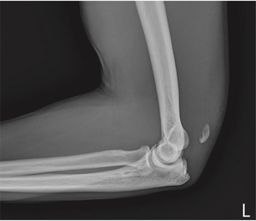
Figure 1: Lateral radiograph of the left elbow of a 39-year-oldmale shows proximal migration of the left patella cubiti withassociated mild overlying soft tissue swelling.
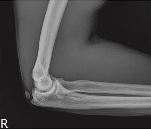
Figure 2: Lateral radiograph of the right elbow of the samepatient shows patella cubiti.
Its etiology is unknown with congenital, developmental ortraumatic injury theories. Kattan2 and others supported acongenital origin of patella cubiti as a sesamoid in the tricepstendon. The developmental theories showed separation of theepiphyseal center of the olecranon in early childhood leadingto independent development of this center.5 According todevelopmental theories by Van Demark6, growth disturbanceoccurring at a particular age could lead to the prevention ofcertain epiphyses of the body from uniting the metaphyses.The traumatic theories of patella cubiti have been reported bymany authors. Habbe7 suggested avulsion of the olecranonepiphysis with periosteal stripping of proximal ulnar diaphysis.O’Donoghue8 reported a case where trauma caused a fracturethrough the cartilaginous epiphyseal growth plate of thepatella cubiti which was surgically fixed, whereas Levine9reported bony fracture through the patella cubiti which wassurgically excised.
The radiology findings of patella cubiti should bedifferentiated from post-traumatic non-union of an old fractureof the olecranon tip. Patella cubiti is well delineated with asmooth intact cortical surface of the olecranon, while nonunionof old fractures should show an irregular surface withsclerotic margin of the parent olecranon and distal fracturefragments. Other differential diagnosis include unfusedolecranon epiphysis in young patients and hydroxyapatitedeposits within the triceps tendon.
A rupture or avulsion of the triceps brachii tendon is a rareinjury, first reported by Partridge10 in 1968. There are manycauses of a ruptured triceps tendon such as traumatic injury,which is usually seen in young, healthy patients and othersystemic diseases such as systemic lupus erythematosus (SLE),rheumatoid arthritis (RA), chronic renal failure, secondaryhyperparathyroidism, hypocalcemic tetany, Marfan syndrome,chronic acidosis, osteogenic imperfecta, and anabolic steroiduse. Other local conditions which were reported to causerupture include xanthoma, hemangiomendothelioma, injectionof corticosteroids, attrition changes from degenerativearthritis and olecranon bursitis.11,12
The study was limited due to no evidence of furtherimaging as there was no follow up of the patient.
Patella cubiti is a patella-like sesamoid bone in the tricepsbrachii tendon. Patients can present asymptomatic orsymptomatic including limitation of motion, stiffness or pain.Radiology features of normal patella cubiti show an ossicleproject posterior to elbow joints. To avoid diagnosis pitfalls,radiologists and clinicians should keep in mind thatassociated proximal migration of ossicles should raise clinicalsuspicion of a ruptured triceps brachii tendon. Suspicion shouldbe heightened when a patient has a known clinical history ofprior injury with abnormal physical examination.


