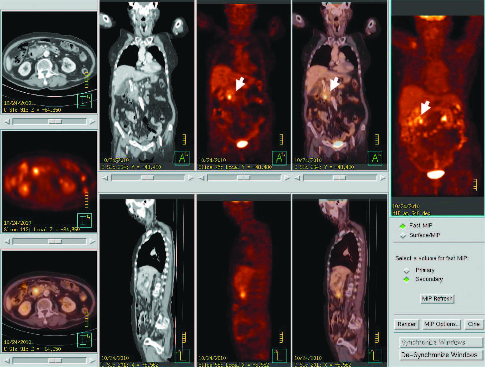
PET/CT images show 1.3 cm. hypermetabolic mass at pancreatic head (arrow), without regional node and distant metastasis (stage T1N0M0)
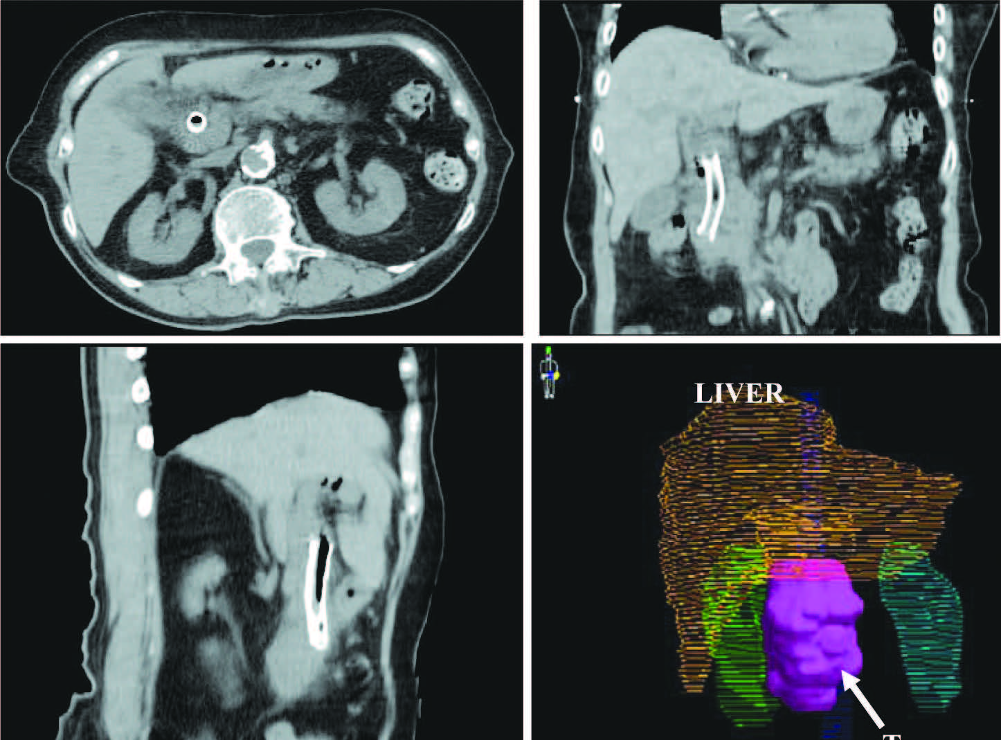
Figure 2:
CT images show the tumor in 3 plane and 3 Dimensional imaging of tumor (pink colour), OAR Liver (yellow), right kidney (green), left kidneys (blue). There is metabolic stent placement in common bile duct.
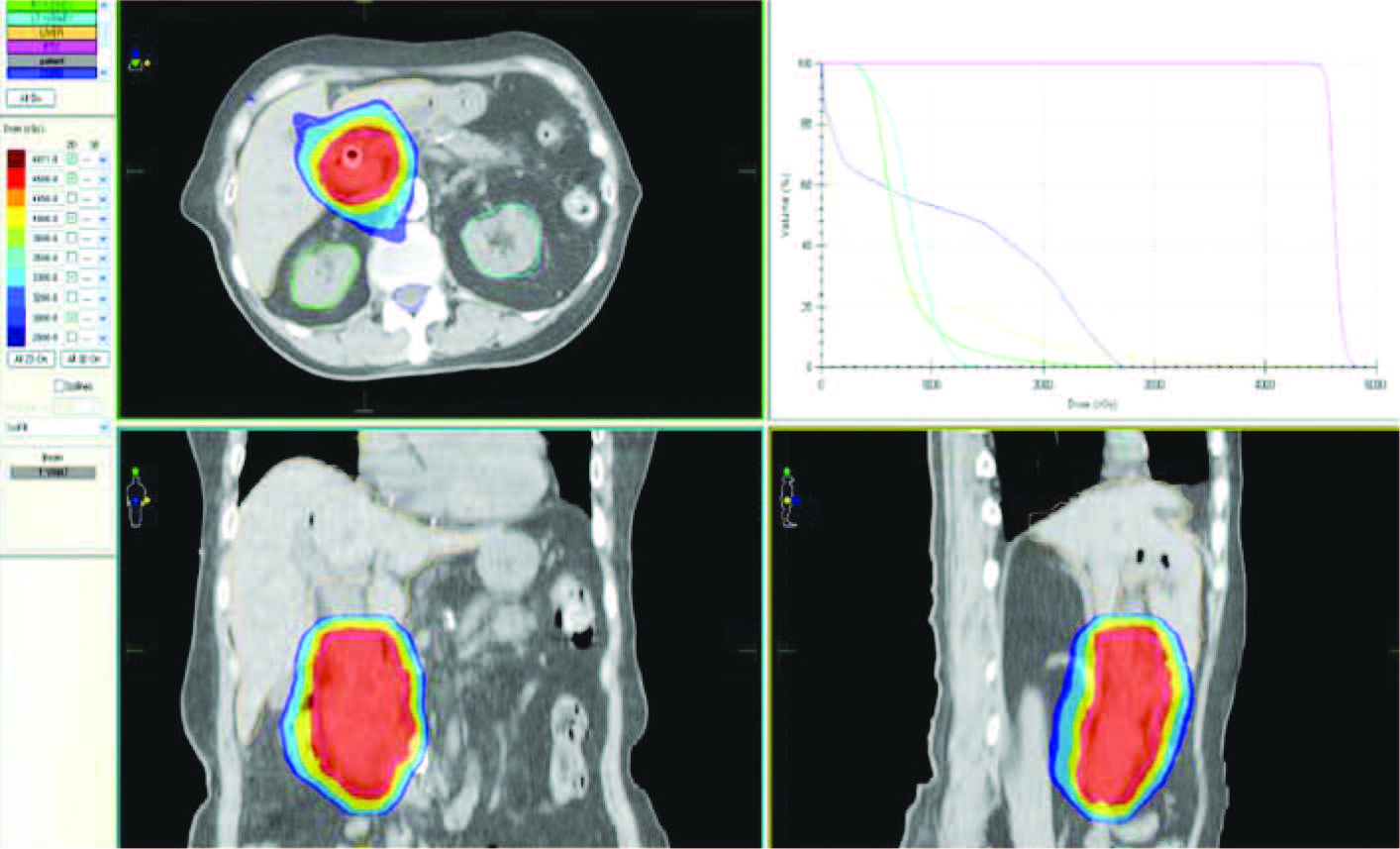
Figure 3:
The 2D distributions for transverse, sagittal, coronal planes and Dose Volume Histograms (DVHs) of VMAT plan
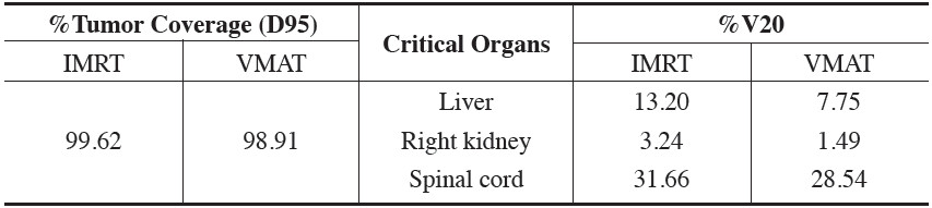
Table 1:
Comparison of IMRT and VMAT plan
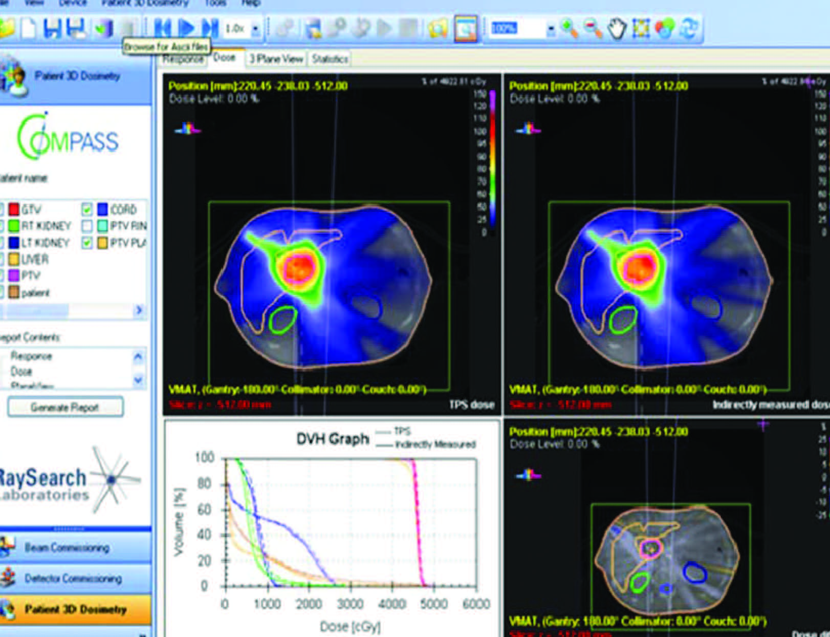
Figure 4:
Shows the result of COMPASS 3D QA for VMAT plan
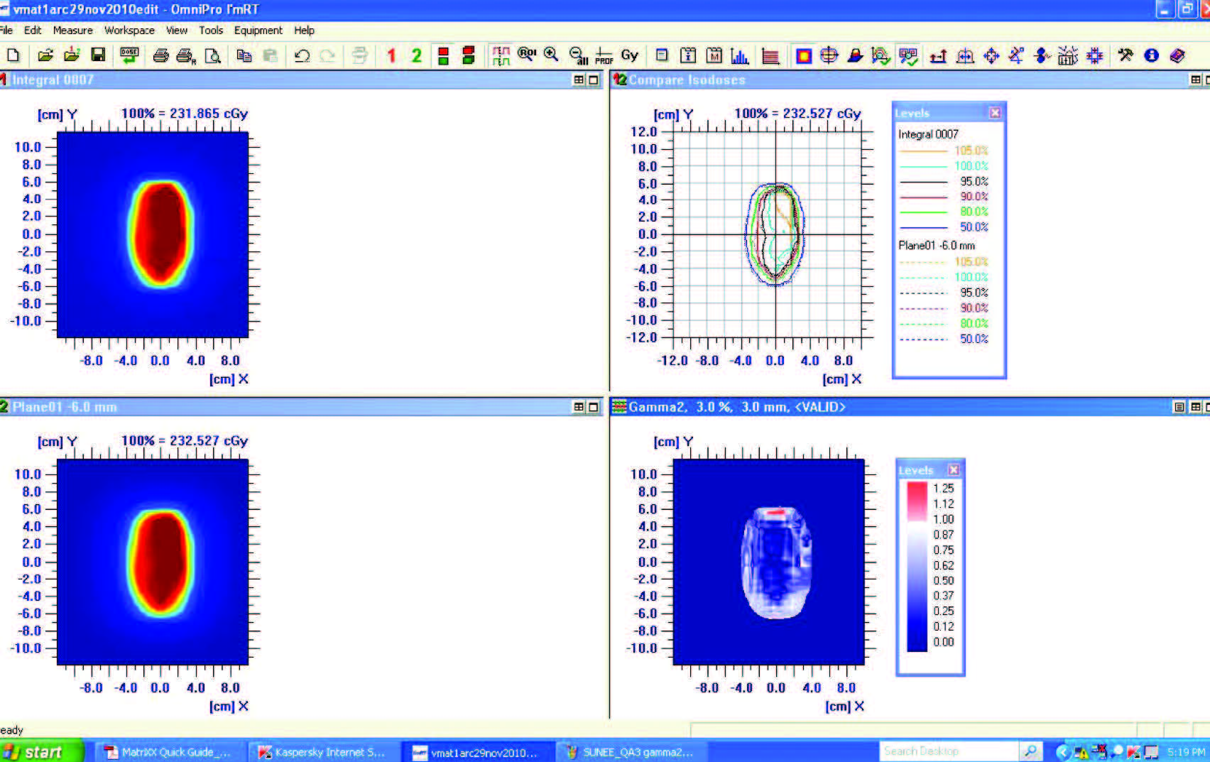
Figure 5:
Shows the result of 2D QA for VMAT plan with Omnipro IMRT
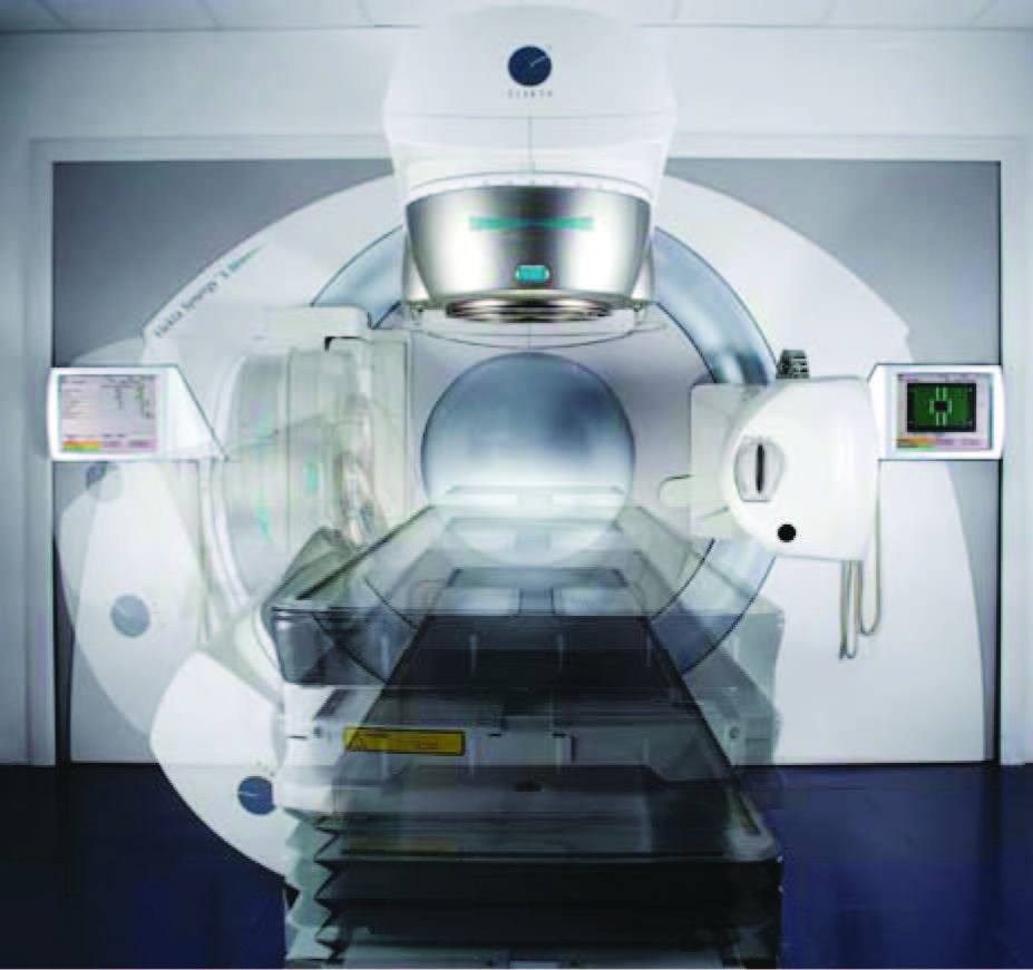
Figure 7:
The Elekta Synergy® combines VMAT&IMRT
and image guided radiation therapy (IGRT), XVI®