Electronic ISSN 2287-0237

VOLUME

PAGET’S DISEASE OF THE NIPPLE: CHOICE OF IMAGING MODALITIES FOR BREAST LOCALIZATION IN CASE OF MULTIPLE BREAST NODULES AND SENTINEL NODE

SEPTEMBER 2015 - VOL.10 | CONTINUING MEDICAL EDUCATION

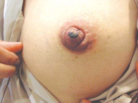
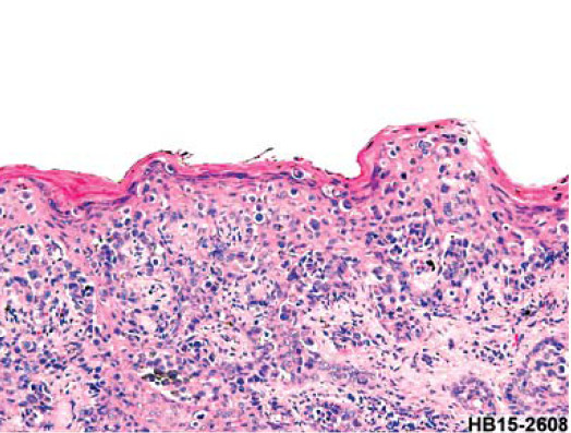
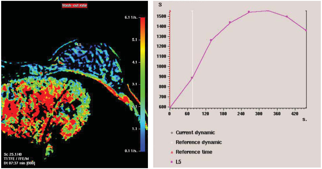
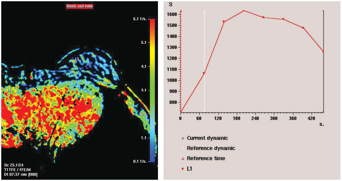
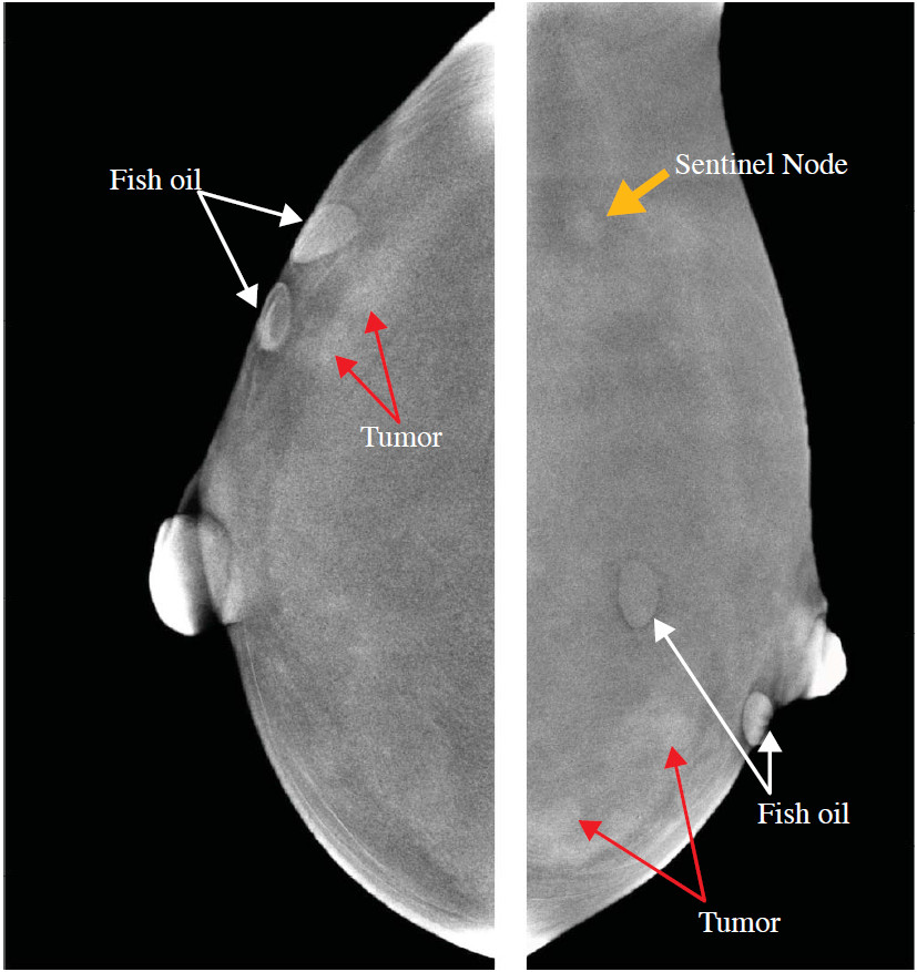
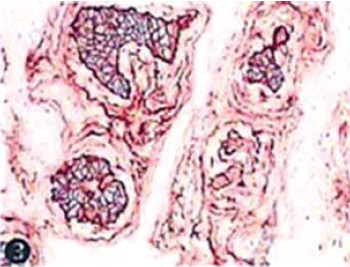
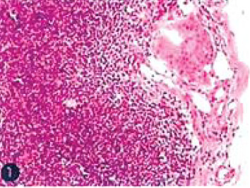
Paget’s disease of nipple is rare entity. The majority of cases are associated with ductal carcinoma in situ (DCIS) or invasive ductal carcinoma (IDC) somewhere in the breast. When ultrasonic scan found multiheterogenic masses in both breasts, it is imposible to vertify which one or more foci to be malignancy. We suggest to mark with small fish oil capsules at suspected masses before magnetic resonance imaging (MRI) breast with contrast enhancement as well as contrast enhanced spectral mammography (CESM) for comparison. The finding shows CESM is far more superior than (MRI) breast study on detection of sentinel node. CESM is faster, inexpensive and far more convenient for the patient.

Keywords

Paget’s disease of the nipple, breast localization, digital mammography, ultrasonic scan, MRI breast contrast enhancement, CESM, sentinel node, contrast enhanced spectral mammography

DOI:

10.31524/bkkmedj.2015.09.014

MEDIA
Figure 1: Localized dark red lesion at left nipple is seen, no ulceration of extension into areolar region is observed.
Figure 2: There are abnormal cells, single units and in groups, in the epidermis, some of them house melanon pigment, no abnormal cells in the dermis.
Immunohistochemical stains CAM5.2 is positive, HMB45 is negative in tumor cells, Melan A is negative in the tumor cells. Paget’s disease of nipple is diagnosed.
Figure 3: MRI of the breasts with dynamic Gd study reveals contrast enhanced masses at the medial aspect of left breast. MRI Gd dynamic study reveals rapidly increased contrast enhancement on the arterial phase
and then this declined gradually on venous and delayed phases (washout) (Figure 4). The findings are suggestive of malignancy. The other masses in either left or right breast are not shown in contrast enhancement.
Figure 4: MRI Gd dynamic study sample on the left breast reveals rapidly increased contrast enhancement on arterial phase and then declined gradually on venous and delayed phases (washout). The findings are suggestive of malignancy.
Only two lesions at the medial aspect of left breast are contrast enhancement, other lesions of both breasts show negative (they probably represent benign fibroadenomas).
Figure 5: Contrast enhanced spectral mammography (with fish oil marker see arrow) on the skin shows small two tumors with contrast enhancement (see arrow). Other lesions are negative for contrast enhancement.
Figure 6A: EGFR -Negative.
Figure 6B1: Estrogen receptor- Positive 60%.
Figure 6B2: Progesterone receptor- Positive 5% (See 2).
Figure 6B3: c-erbB2 (HER2) Score 3+ = Positive (Circumferential membrane staining that is complete, intense, and within > 10% of tumor cells).
Figure 6C: Sections of sentinel lymph node shows tumor cells (< 2mm) in one lymph node.
SEARCH VOLUME & NUMBER
GO
RECENT VOLUME & NUMBER