Electronic ISSN 2287-0237
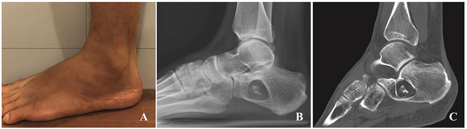
A 32-year-old Cambodian malepresented with intermittentright ankle pain withouthistory of trauma. Physical examinationrevealed normal configuration withoutswelling, mild tenderness at anterolateraland anterior (A). Lateral view of rightankle radiograph demonstrated awell-defined osteolytic lesion at rightcalcaneal neck to body, with thinsclerotic rim, containing focal internal dystrophic calcification, without adjacent cortical breakthrough (B). There is a diffuse fat density in the lesion on non-contrast
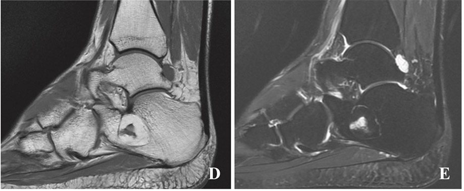
computed tomography (CT) (C), corresponding to additional magnetic resonance imaging (MRI) findings; bright T1W signal
(D) with signal drop on fat suppression sequence, and small surrounding vascularity (E). Intraosseous calcaneal lipoma was
diagnosed.
The fat component is the key of imaging findings. CT and MRI are diagnostic modalities, able to demonstrable fat component;fat density (-60 to 100 hounsfield unit-HU) on CT, bright T1W and T2W signals as subcutaneous fat with signal drop on fatsuppression sequences, but mildly brighter signal than fatty marrow, due to cellularity of marrow. Bone expansile remodelingcould be also seen.1
Intraosseous lipoma is very rare benign bone tumor, approximately 0.1 to 2.5% of all benign bone tumors.2 The firstreported case of intraosseous lipoma was in 1910. Most of the patients with intraosseous lipoma are asymptomatic and the bonetumor is found incidentally.2 Some authors believe that tumor associated with pain. The tumor can occur in all age groups, butis usually found at the age of 40, and more observed in males than females.1,2 Normally there is no treatment for asymptomaticpatients with intraosseous lipoma, some can evolve as extensive fat necrosis, calcification and cystic change, but if there is somesuspicion of malignancy, tissue diagnosis and surgery treatment is available.1-3


