Electronic ISSN 2287-0237

VOLUME

DETECTION OF CORONARY BYPASS GRAFT PATENCY BY 256-SLICE MULTI-DETECTOR COMPUTED TOMOGRAPHY

SEPTEMBER 2011 - VOL.2 | ORIGINAL ARTICLE
OBJECTIVE

To evaluate the diagnostic accuracy of 256-slice Multidetector Computerized Tomography (256-MDCT) in detection of coronary graft patency by comparison with the gold standard invasive coronary angiography (ICA).

MATERIALS AND METHODS

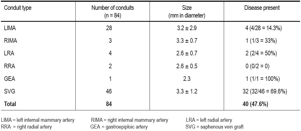
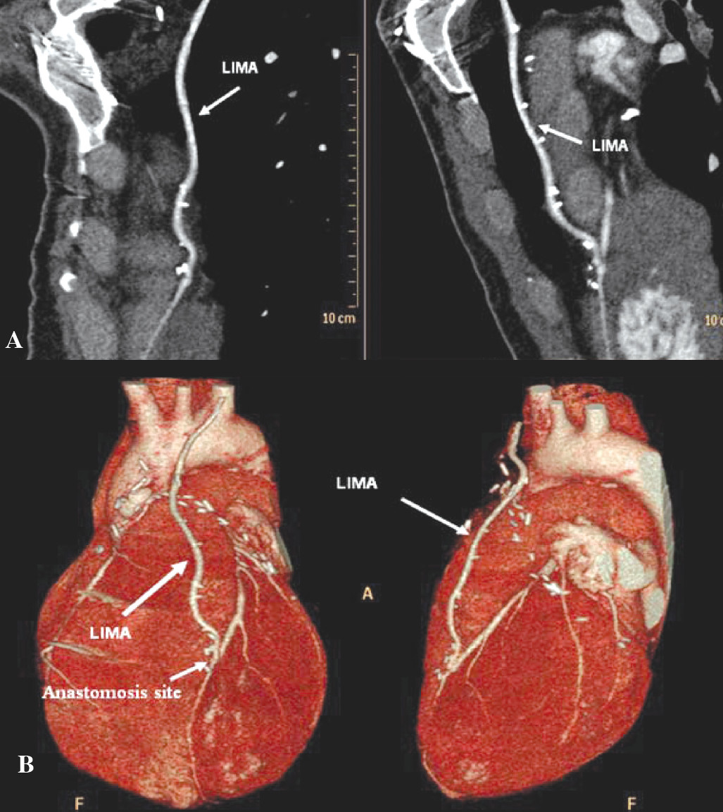
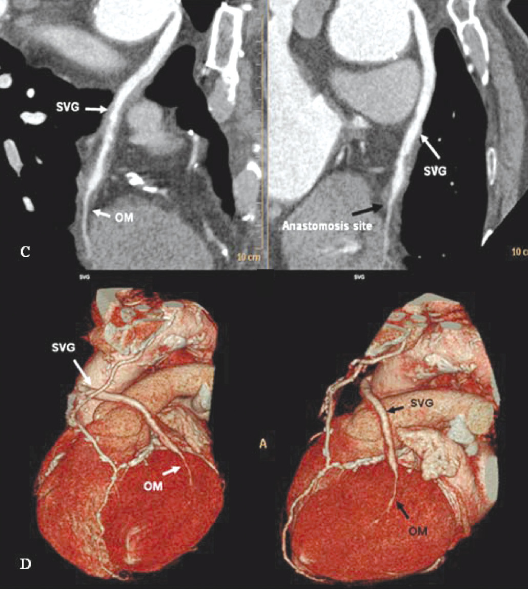
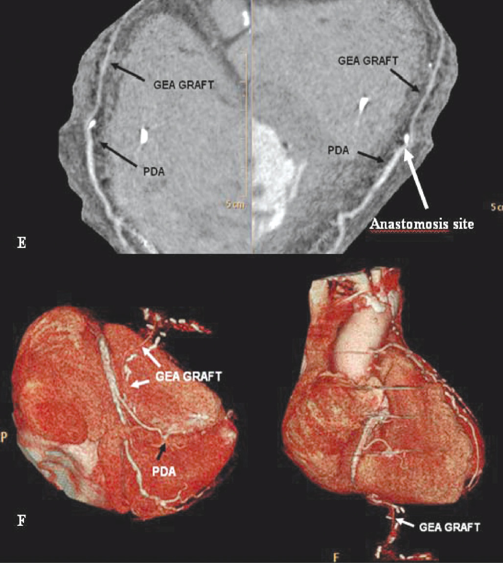
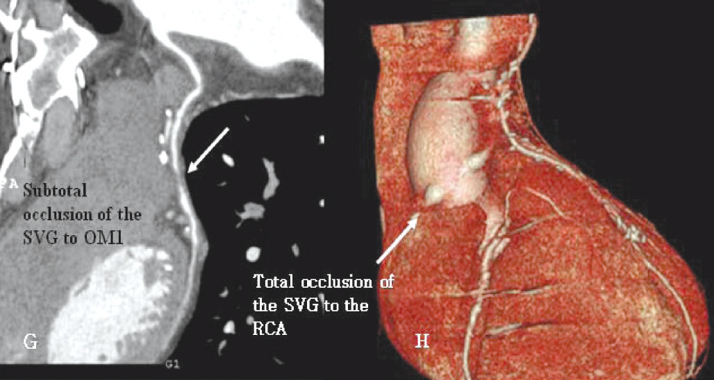
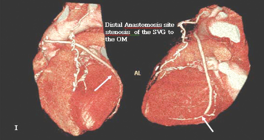
From January 2009 to April 2011, a total of 29 consecutive patients who had previously had CABG surgery were referred to us for assessment of graft patency. A total of 84 coronary bypass graft conduits (38 arterial graft conduits, 46 venous graft conduits) were studied, using 256-MDCT and ICA with iodine contrast intravenous injection. All patients underwent coronary angiography to either confirm result or PCI of graft disease. The diagnostic accuracy of the 256-MDCT for coronary bypass graft evaluation was assessed by comparing it to the ICA in terms of sensitivity, specificity, positive predictive value (PPV) and negative predictive value (NPV).

RESULTS

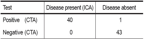
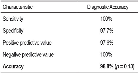
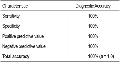
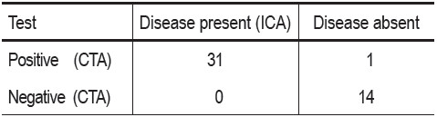
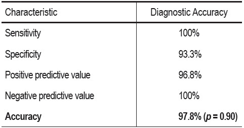
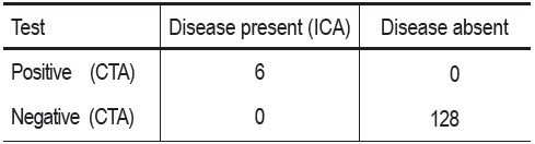
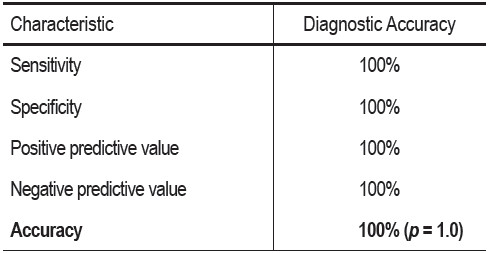
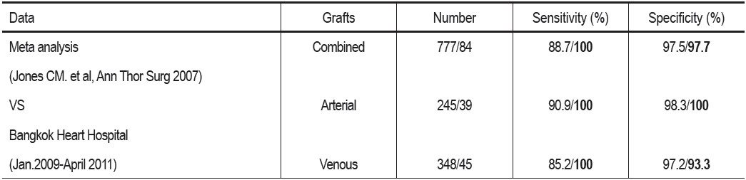
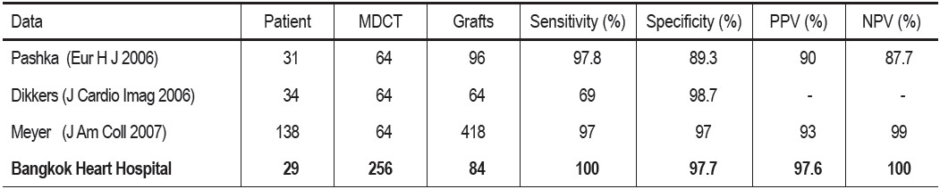
A total of 84 grafts were imaged using the 256-MDCT and all grafts were visualized. There was no statistical difference in diagnostic accuracy between MDCT and ICA regardless of the age, size or type of the bypass graft conduit (p value = 0.13). The sensitivity, specificity, positive predictive value, negative predictive value of 256-MDCT in coronary artery bypass graft assessment were 100%, 97.8% , 97.5% and 100 % respectively.

CONCLUSION

The 256-MDCT provides a high accuracy, reliability and feasibility for coronary bypass graft evaluation and the diagnostic accuracy is comparable to the gold standard ICA.

Keywords

Coronary bypass graft, 256-MDCT, Invasive coronary angiography, Sensitivity, Specificity, Positive predictive value, Negative predictive value, Accuracy

DOI

10.31524/bkkmedj.2011.09.002

MEDIA
Table 1: Coronary bypass graft conduit characters.
Table 2: Correlative findings of 84 coronary bypass graft conduits between CT coronary angiography (CTA) and invasive coronary angiography (ICA).
Table 3: Overall diagnostic accuracy of the 256-MDCT compared to the gold standard invasive coronary angiography (ICA).
Table 4: Correlative findings of 38 arterial bypass graft conduits between CT coronary angiography (CTA) and invasive coronary angiography (ICA).
Table 5: Overall diagnostic accuracy of the 256-MDCT compared to the gold standard invasive coronary angiography (ICA) in arterial bypass graft assessment.
Table 6: Correlative findings of 46 venous bypass graft conduits between CT coronary angiography (CTA) and invasive coronary angiography (ICA).
Table 7: Overall diagnostic accuracy of the 256-MDCT compared to the gold standard invasive coronary angiography (ICA) in coronary venous bypass graft assessment.
Table 8: Overall diagnostic accuracy of the 256-MDCT compared to the gold standard invasive coronary angiography (ICA) in 134 anastomosis sites of the coronary bypass graft assessment.
Table 9: Overall diagnostic accuracy of the 256-MDCT compared to the gold standard invasive coronary angiography (ICA) in 134 anastomosis sites of the coronary bypass graft assessment.
Table 10: Comparison of the overall diagnostic accuracy of the 256-MDCT to the gold standard invasive coronary angiography (ICA) in bypass graft assessment to the meta analysis studies of the 16-, 64-MDCT.
Table 11: Comparison between the accuracy of the 256-MDCT (Bangkok Heart Hospital) with 64-MDCT in bypass graft assessment studies of the others.
Figure 1: Demonstration of LIMA graft to the LAD and its anastomosis site.
A: MPR images. B: Volume rendering images. LIMA = left internal mammary artery, LAD = left anterior descemding, MPR = multiplanar reconstruction
Figure 2: Demonstration of SVG to the OM and its anastomosis site.
C: MPR images shows the distal stenosis of the SVG. D: Volume rendering images. SVG = saphenous vein graft, OM = obtuse marginal branch, MPR = multiplanar reconstruction
Figure 3: Demonstration of the GEA to the PDA and its anastomosis site.
E: MPR images shows the patent of the GEA graft. F: Volume rendering images. GEA = Gastroepiploic arterial graft, PDA = Posterior descending artery, MPR = multiplanar reconstruction
Figure 4: Demonstration of graft vessel stenosis.
G: MPR images. H: Volume rendering image. MPR = multiplanar reconstruction, SVG = saphenous vein graft, OM = obtuse marginal branch, RCA = right coronary artery
Figure 5: Demonstration of distal anastomosis site stenosis of the SVG to the OM.
SVG = saphenous vein graft, OM = obtuse marginal branch
SEARCH VOLUME & NUMBER
GO
RECENT VOLUME & NUMBER