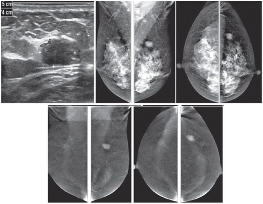
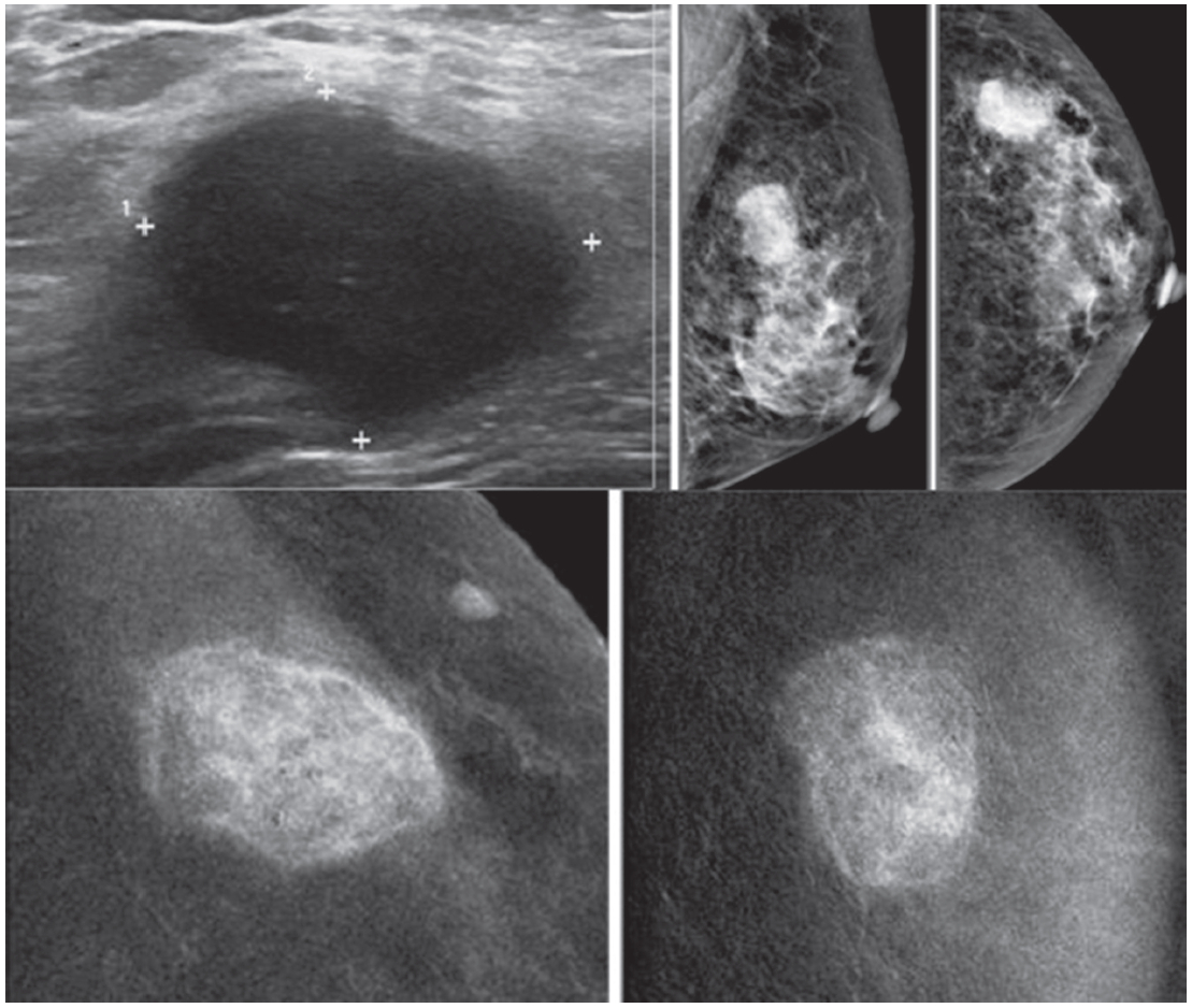
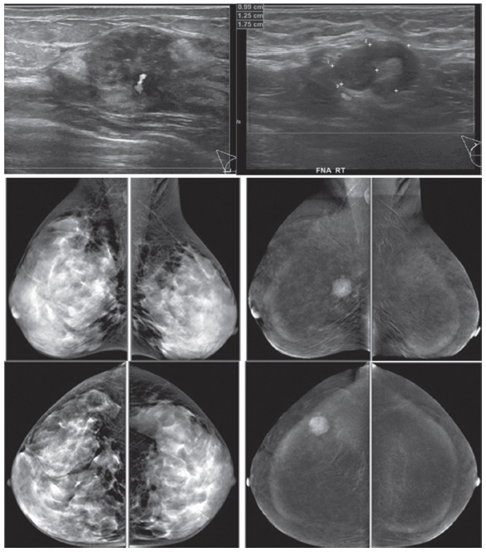
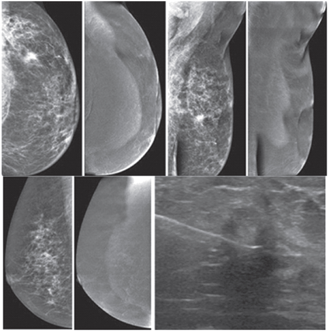
Figure 1A:
Female 48-years of age, ultrasound (US) shows a microlobulate, irregular shaped cystic mass, with internal solid component. Multiple enlarged axillaries denote a denopathy with round shape, almost echo-free, no fatty hila are seen. Mammography shows focal density in craniocaudal (CC) view, not well defined in mediolateral oblique (MLO) view. CESM shows intense enhancement of that complex lesion, with its size much increased. Numerous enhanced small foci are noted in both breasts. US guided core needle biopsy (CNB) reveals invasive ductal carcinomas and these are confirmed in surgery.
Figure 1B:
Female 42-years of age, US shows a relatively large homogeneoushypoechoic solid mass in a cystic lesion with relationship to ducts, measuring 5.2×7.2mm in right upper outer quadrant (UOQ) and 7.3×11.2mm in left UOQ. Mammography shows a density suspected in left MLO at a second look. CESM reveals a moderate degree of uptake of contrast medium in left UOQ, measuring 11.6×11.9mm and an ill-defined focal minimal abnormal uptake in the right breast. CNB reveals fibrocystic changes of both lesions.
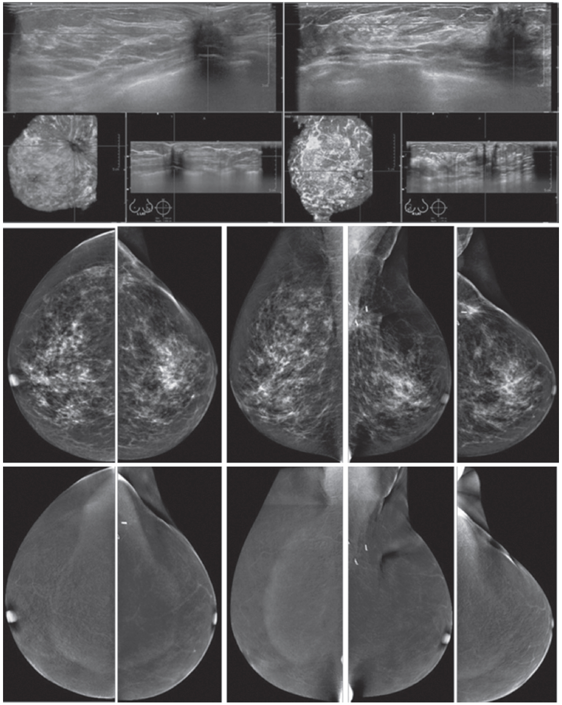
Figure 1C:
Female 64-years of age, post op left breast conserving therapy (BCT). Mammography shows architectural distortion at surgical scar in left lower outer quadrant (LOQ). US reveal an enlarging cystic lesion with hypoechoic modularity on its wall and some echoic contents. CESM shows no enhancement of either breast. US guided aspiration yields old hemorrhagic fluid. At surgery, there are no residual malignant cells.
Figure 1D:
Female 60-years of age, US shows a well-defined lobulated, minimally heterogeneous hypo-echogenic mass with increased vascularity in right LOQ of 2×1.2×2cm. Mammography shows coarse pleomorphic microcalcifications in the areas of the obscured masses. Axillary nodes are seen in the right MLO. CESM reveals an intensely heterogeneous and enhanced microlobulated mass in right LOQ, measuring 18×22mm, with multiple dark spots of non-enhanced microcalcifications inside the lesion. Multiple foci of mild and moderate enhancement are seen. Soft enhanced right axillary nodes are noted. CNB and surgical pathology reveal invasive ductal carcinoma.
Figure 1E:
Female 46-years of age, US shows a well-defined lobulated, minimally heterogeneous hypo-echogenic mass with increased vascularity in the left UOQ. Mammography shows a lobulated isodensity mass. CESM reveals a markedly homogenous enhanced lobulated mass. The outline is smooth and no spiculation is noted. CNB reveals a fibroadenoma.
Figure 1F:
Female 42-years of age, US shows a well-defined round to oval shaped, minimally heterogeneous hypo-echogenic mass with increased vascularity in the right subareolar area. Mammography shows a round isodense mass in the right SA in CC, partially obscured in MLO. CESM reveals no enhancement of this subareolar lesion a benign finding. CNB reveals fibrocystic changes.
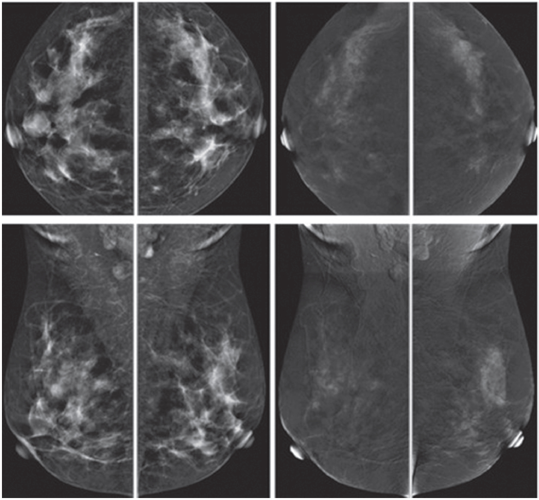
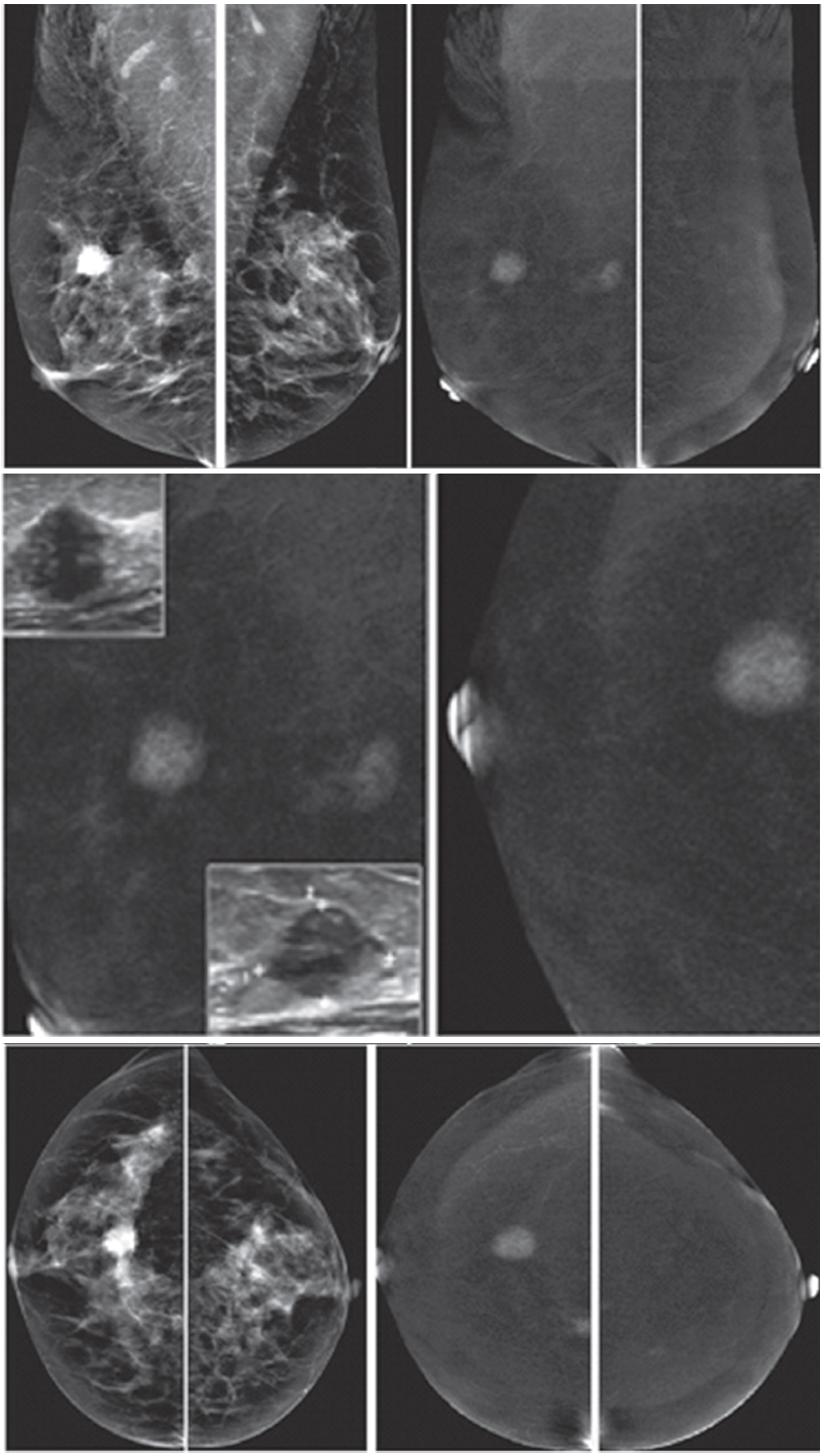
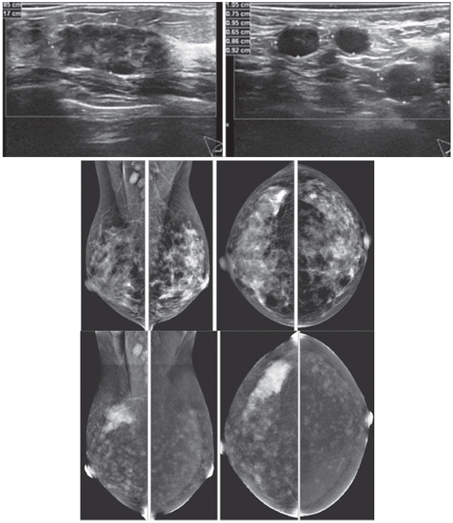
Figure 1G:
Female 52-years of age, US of both SA areas show the same findings of a very low hypoechoic lesion with its depth more than width. Acoustic shadowing and extensive spiculation are seen. This may be seen in malignancy and benign lesions such as radial scar or sclerosingadenosis. However the former lesion is enhanced, while the 2 latter lesions show no significant enhancement. Mammography shows extensive breast asymmetry, irregular shaped lesion with extensive spiculation. CESM reveals very high uptake of contrast medium in almost the whole fibroglandular tissue in both breasts, with long spiculations to nipple and skin on a deep to posterior aspects, compatible with extensive involvement of malignancy. CNB reveals bilateral extensively invasive ductal carcinoma, grade II.
Figure 1H:
Female 34-years of age, the automated breast volume scanner (ABVS) shows 2 poorly defined focal hypoechoic areas in left UIQ and right upper, associated with disruption of parenchyma and a lobulated hypoechoic mass in right UOQ: 7×9.9×12.6mm Mammography reveals architectural distortion in both areas, seen with no mass in a six category classification (SCC). CESM reveals multiple focal enhanced nodules of varying degrees, scattered in both breasts. Pathological study reveals a fibroadenoma with sclerosingadenosis.
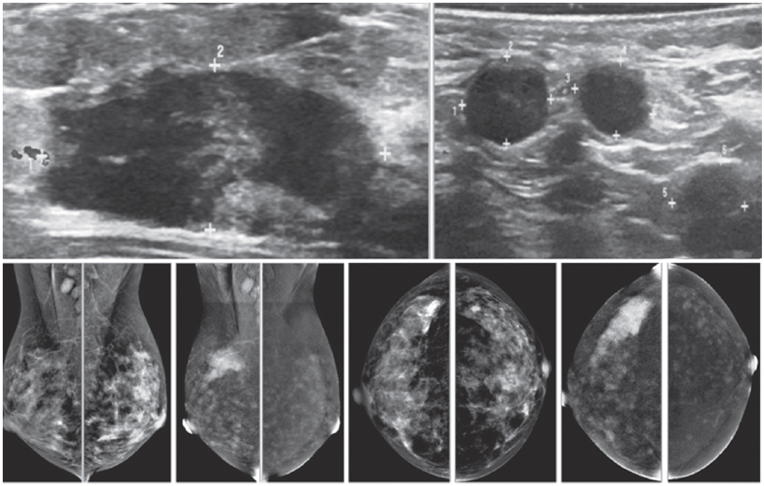
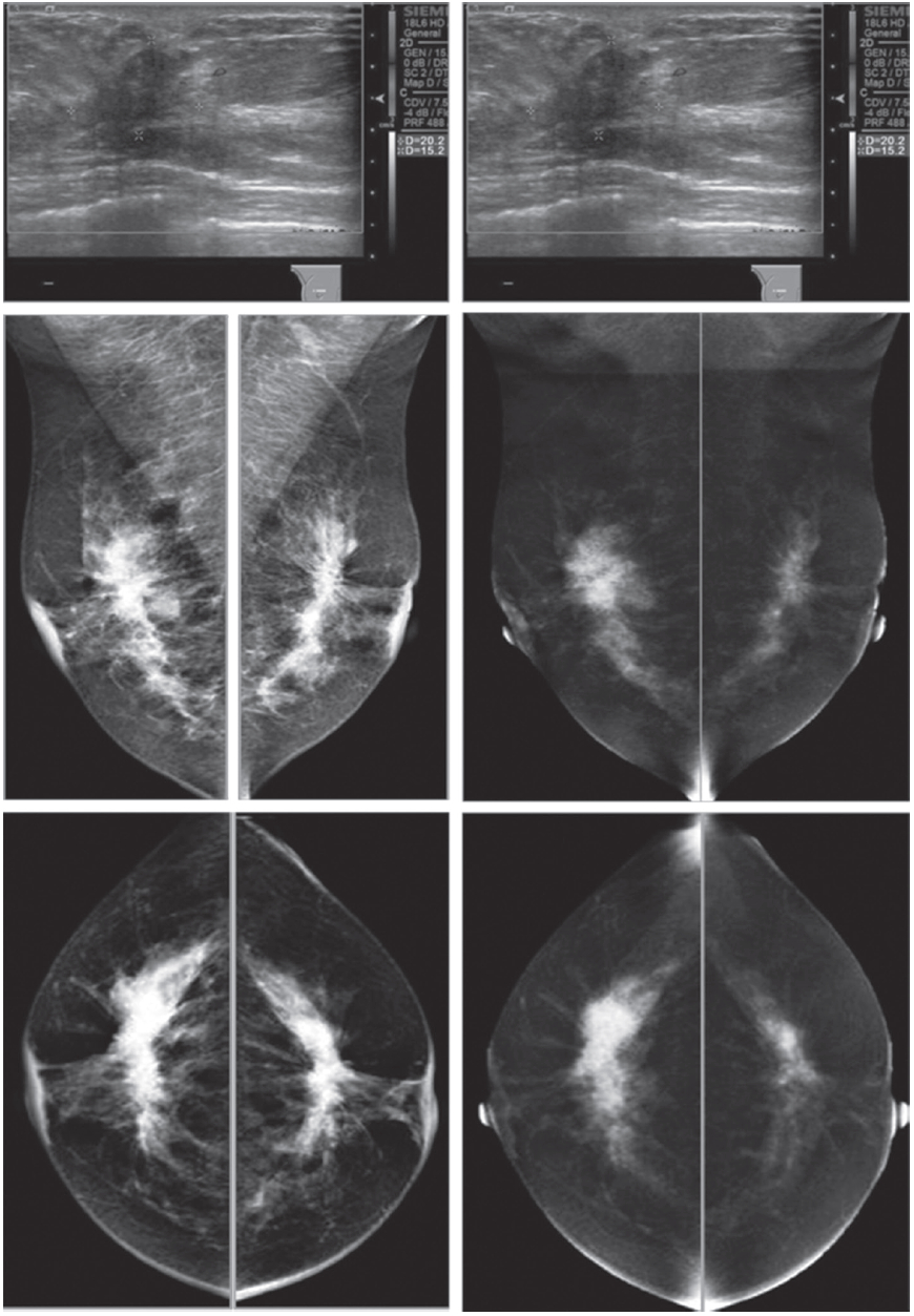
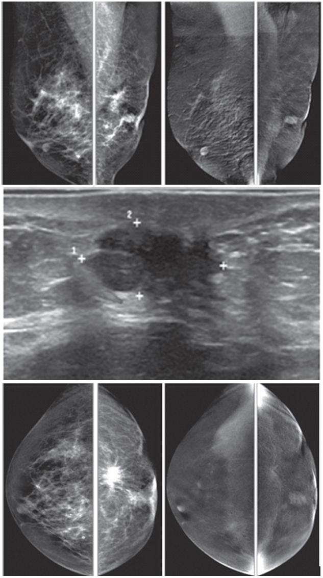
Figure 2A:
Female 60-years of age, a mass is palpable in left UOQ Mammography shows a lobular shaped hyperdense mass and
US shows an echo-free lobular mass with surrounding tissue reaction, combined type of posterior enhancement. CESM reveals an intensely heterogeneous enhancement of a microlobulate mass of 33×24×22mm in right UOQ, seen with multiple dark spots of non-enhanced microcalcifications inside the lesion, highly suggestive of malignancy. Another small markedly enhanced nodule is seen, which is not noticed initially by mammography and US. There is no abnormal enhancement of the entire right breast and axillary l.n. CESM defines the nature and existing of two cancers in the same breast.
Figure 2B:
Female 60-years of age, S/P left CBS, a round mass with microlobulation and spiculation is seen in right upper. Multiple
equivocal nodes are seen in both axillae. CESM reveals aheterogeneous uptake of contrast medium in right upper of 16×17mm. An additional lesion is seen in right inner: 9×11mm. No abnormal uptake in left breast and axillary l.n. Second look US reveals two heterogeneous hypoechoic masses with microlobulated outlines. US guided CNB reveals invasive ductal carcinoma of both lesions. CESM detects the second primary multifocal cancers in contralateral breast.
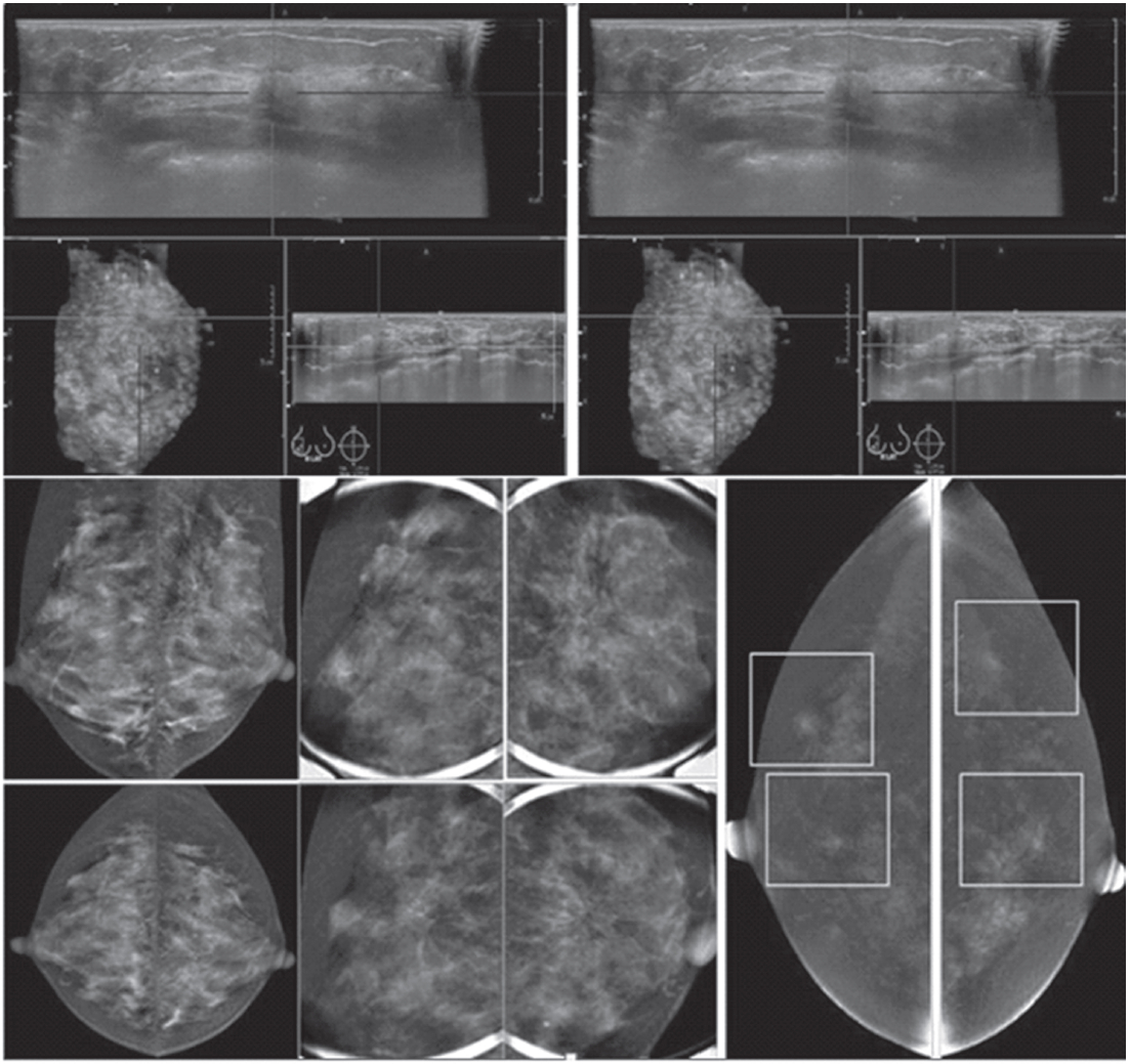
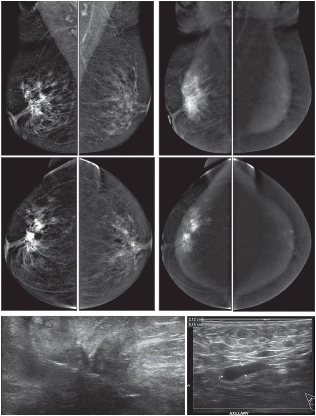
Figure 2C:
Female 55-years of age, mammography shows a large irregular shaped focal asymmetry with spiculation in right breast.
Ultrasound shows a large spiculated mass. The number of small axillary nodes has increased. CESM reveals an intensely heterogeneous enhancement of a large irregular-shaped mass with multiple dark spots of non-enhanced microcalcifications inside the lesion. Spiculations are seen around the lesion; extending anteriorly to skin and nipple and posteriorly to deep structures. Soft enhanced foci in both breasts and left axillary l.n. are noted, highly suggestive of malignancy. US guided CNB reveals invasive ductal carcinoma.
CESM defines the nature, local extension and existence of this cancer, possible bilaterally.
Figure 2D:
Female 50-years of age, mammography and ABVS reveal a malignant appearing mass under 2 cm. in right UOQ, however, multiple enlarged adenopathy is noted. CESM shows the malignant nature of the mass as well as enhancement of multiple axillary adenopathy. Apart from that, another enhanced focus is seen in left breast. A tiny left axillary l.n. is minimally enhanced. Surgical pathology confirms invasive ductal carcinoma grade II with l.n. metastasis in 12 out of 18 removed nodes. CESM defines the nature of this small cancer, but with extensive adenopathy and possible additional small lesion in contralateral breast.
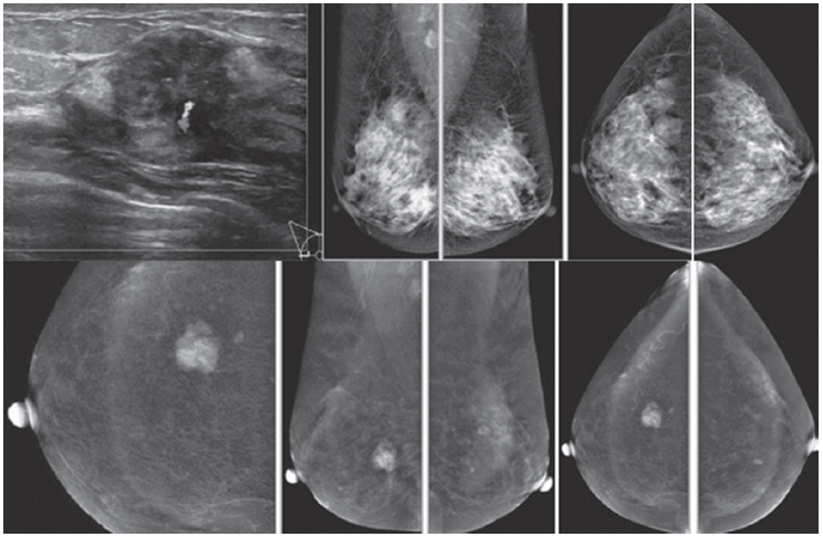
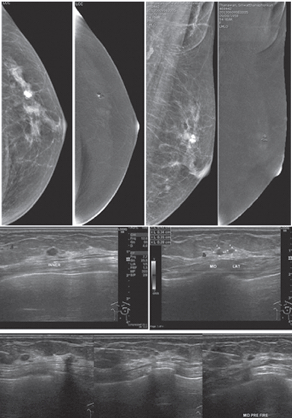
Figure 3:
Female 45-years of age, screening in patient with family history of breast cancer. Mammography shows extremely dense breast, no lesion was detected initially. US reveals a round heterogeneously hypoechoic mass in right outer with abnormal vessel inside the lesion. A round axillary l.n. is seen with increased cortical thickness and focal bulging of the cortex, compatible with micrometastasis. CESM reveals an intensely, heterogenous enhanced round mass of 16x19x20.5mm., with partially seen enhanced axillary l.n. US guided CNB reveals invasive ductal carcinoma. CESM demonstrates a cancer with axillary adenopathy in a high risk patient, while the clinical examination and mammography is negative.
Figure 4:
Female 48-years of age presents with palpable right axillary l.n. US confirms numerous axillary adenopathy and a welldefined mixed echoic mass in right UOQ. Mammography shows a focal density in right CC, obscured in the first look at right MLO. CESM reveals an intensely heterogenous enhancement of a large irregular shaped mass, seen with some dark spots of non-enhanced microcalcifications inside the lesion. Enormous moderately enhanced foci are seen in both breasts. The study was performed near the menstruation, thus repeat study should be performed to differentiate extensive bilateral cancer or physiological enhanced foci due to hormone effect. The partially seen enhanced right axillary nodes are extensive. CESM detects and confirms the unconvincing
mammography abnormality, axillary adenopathy and possible extensive bilateral lesions or hormonal effect.
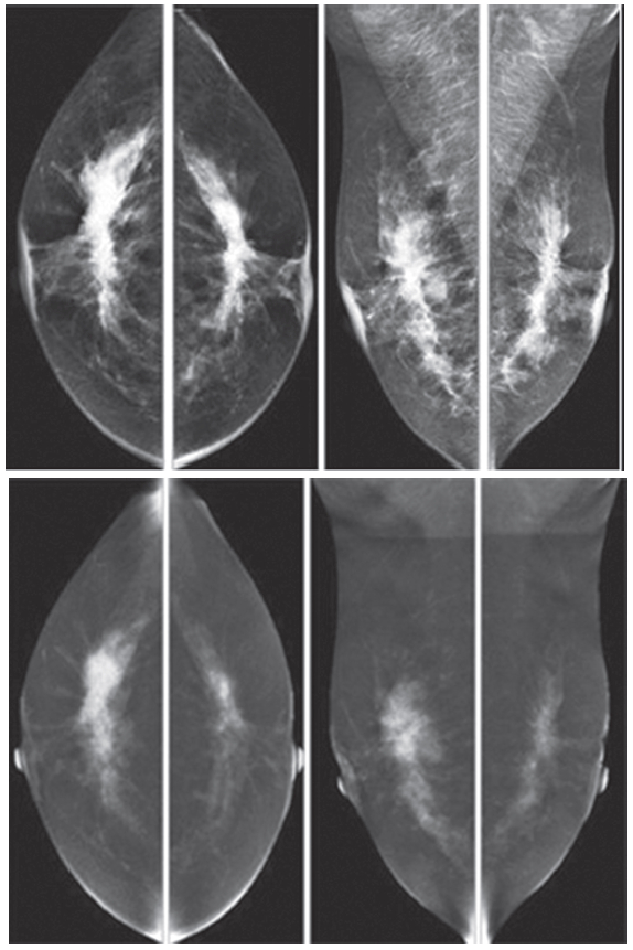
Figure 5:
Female 52-years of age, CESM reveals very high uptake of contrast medium in almost the whole fibroglandular tissue
in both breasts, with long spiculation to nipple, skin and deep to posterior aspects. CNB reveals bilateral extensive invasive ductal
carcinoma; grade II, not suitable for surgery. Chemotherapy was given and CESM will be performed to evaluate the results of the
treatment.
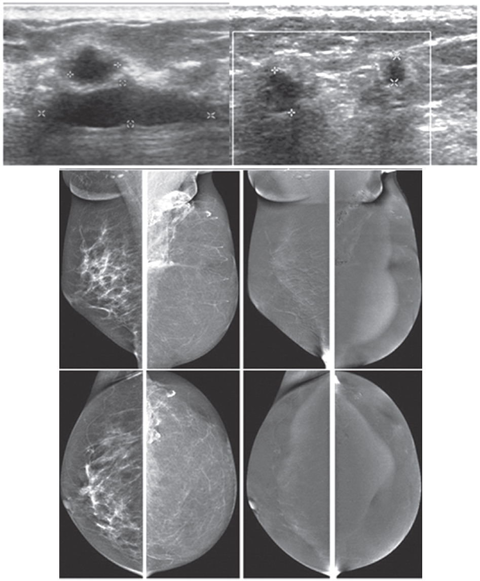
Figure 6A:
Female 51-years of age, S/P left MRM with TRAM flap for left breast cancer. Mammography shows dystrophic calcified areas in far left UOQ US shows 9 lobulated echo-free lesions with wall thickening. There is no typical posterior enhancement that is usually seen in cystic lesions. CESM reveals no significant enhancement of both breasts, including at the dystrophic calcified area, compatible with oil cysts post TRAM flap, no evidence of recurrence. CESM post TRAM flap operation excludes local recurrent in
multiple oil cysts with dystrophic calcifications.
Figure 6B:
Female 60-years of age, S/P left MRM with tram flap. Mammography shows a small spiculated mass in PO area, noted with other PO changes. CESM: No abnormal enhancement in both breasts, over all non- malignant lesions. U/S guided CNB of an irregular-shaped markedly hypoechoic mass reveals no residual cancer. CESM post TRAM flap operation excludes local recurrent in the PO spiculated mass.
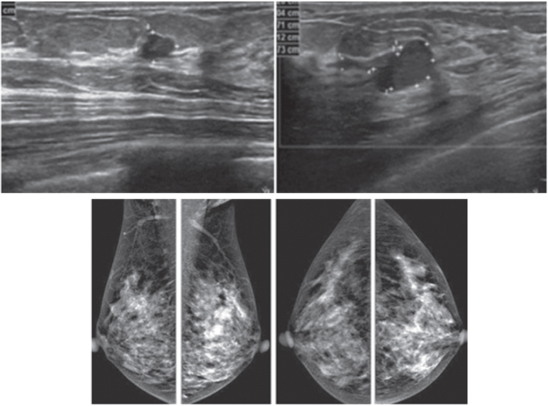
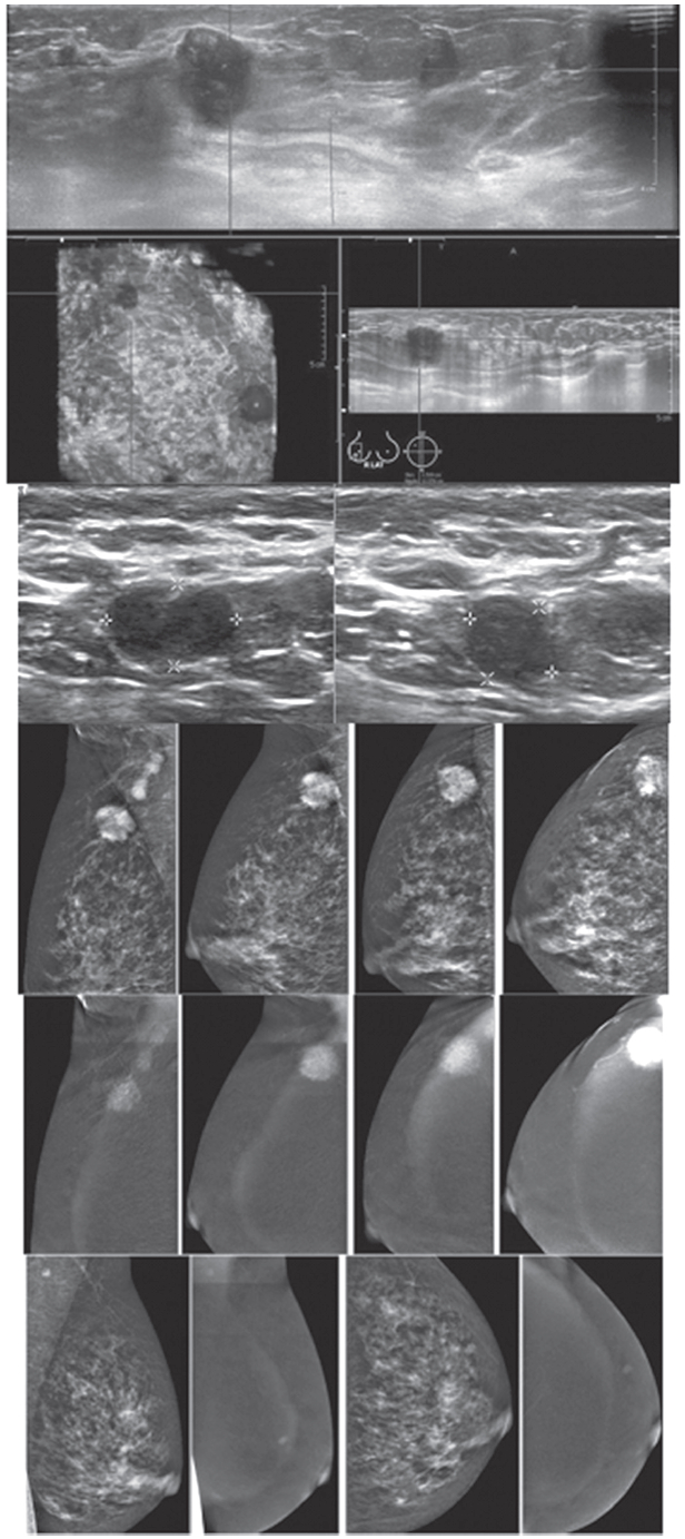
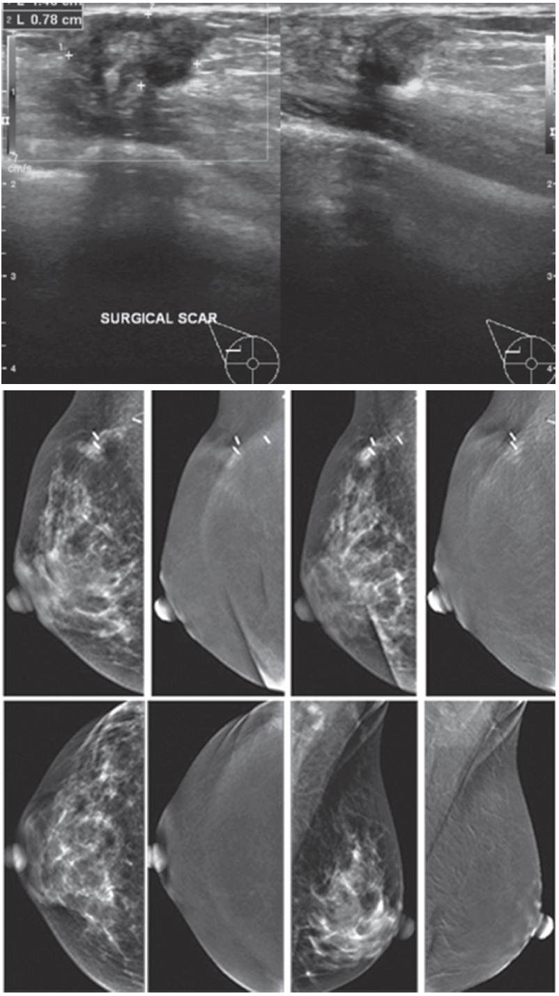
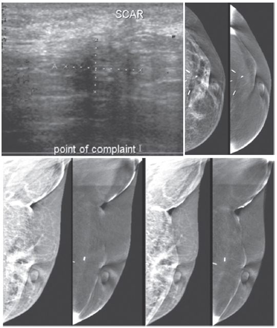
Figure 7A:
Female 54-years of age, S/P lumpectomy in the left upper quadrant noted with palpable abnormality in PO area, not at the left subaredar SA. US shows an irregular mass-like lesion with spiculation in left central at post operation scar. A well-defined heterogeneous hypoechoic lobulated mass is noted in left SA: 8.1 x 16.2 mm CESM reveals the irregular mass-like lesion with spiculation in left central is not enhanced while the non-palpable nodule is densely enhanced in heterogeneous pattern. (The skin mole in right LOQ is seen with enhancement.) At surgery, the post-operative scar shows no malignancy, while the non-palpable SA mass is a recurrent invasive ductal carcinoma. CESM confirms no local recurrence, but another recurrent cancer is noted, slightly away from the PO scar.
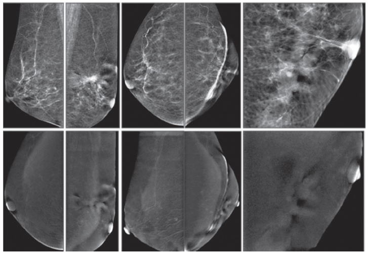
Figure 7B:
Female 54-years of age, S/P left MRM for IDCA. Mammography reveals popcorn calcifications and a cluster of round microcalcifications in left UOQ. Breast US shows 3 microlobulated heterogeneous hypoechoic masses in left upper inner: 4.3×4.9×5mm, left upper middle: 3.1×4.3mm and left upper lateral: 3.1×4.5mm. CESM reveals very soft enhancement of these 3 tiny nodules. US guided CNB of these 3 lesions reveal IDCA, moderately differentiated in all specimens.
Figure 7C:
Female 36-years of age, S/P right CBS for breast cancer came with palpable abnormality at PO area. US shows a heterogeneous hypoechoic mass, lobulated outline, near PO scar, 14.0x7.8 mm. with multiple heterogenous hypoechoic masses in both breasts, and recurrence cannot be excluded. CESM reveals soft enhancement at the PO area with palpable abnormality and surgical clips. There is no significant abnormal enhancement in the rest of both breasts. At surgery, there is no local recurrence. CESM confirms no local recurrence in equivocal mammography and US findings, as well as no evidence of malignancy elsewhere.
Figure 7D:
Female 46-years of age, S/P left UOQ lumpectomy and left sentinel node biopsy for IDCA, grade II US shows an irregular mass--a lesion with spiculation in left central at post operation (PO) scar. Mammography shows architectural distortion at surgical scar with surgical clips in left UOQ.
Figure 7E:
Female 56-years of age, S/P left BCT for invasive ductal carcinoma, came with palpable abnormality in left axilla. US shows an ill-defined abnormal echoic area, no confined mass in left axillary area, at the palpable abnormality, measures 11×16mm. Mammography shows PO changes, no associated mass.
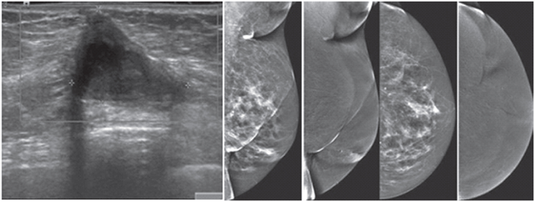
Figure 7F:
Female 74-years of age, mammography shows a spiculated mass in PO area with severe deformity, skin thickening and thick strand extends to the nipple. CESM shows no enhancement of the mentioned areas. No local recurrence is detected. However, a few tiny soft enhanced foci are seen in both breasts, and this requires close follow-up. CESM confirms no local recurrence at PO scar and but cannot exclude tiny foci of malignancy, and this requires follow-up.