Electronic ISSN 2287-0237
To assess the changing of pelvic incidence in patients who received lumbar and lumbosacral fusion with pedicle screw fixation.
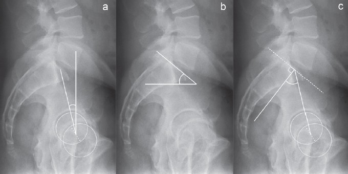
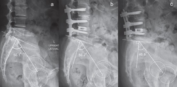
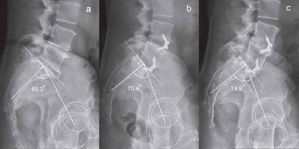
This is a single-institute, retrospective study. From 2011-2016, 113 cases of lumbar and lumbosacral fusion with pedicle screw fixation were reviewed. Preoperative and postoperative (at 6-week, 1-year and latest follow-up) pelvic parameters including Pelvic incidence (PI), Pelvic tilt (PT), Sacral slope (SS) and Lumbar lordosis (LL) were measured in standing lateral view X-ray by 2 independent fellow-trained spine surgeon who were blind to the operation. Difference in preoperative and postoperative PI was defined as Pelvic incidence disparity (ΔPI). Other characteristic data of patients were also collected, including age, sex, body mass index, diagnosis, fusion technique, number of fusion levels and level of fusion.
Pelvic incidence disparity (ΔPI) was 3.2º ± 4.0 at 6-week postoperative, 3.3º ± 4.0 at 1-year postoperative and 3.2º ± 3.4 at last follow-up. This showed a significant change when compared to preoperative but did not change significantly over time after surgery. There was no correlation between ΔPI and fusion technique, L5-S1 fusion, diagnosis and number of fusion segments.
Lumbar and lumbosacral fusion with pedicle screw fixation can alter pelvic incidence parameters. This could be a consequence from increased stress and motion in SI joint after the surgery.
pelvic incidence, spinopelvic parameter, lumbar fusion, sacroiliac joint
Received: May 13, 2018
Revision received: May 21, 2018
Accepted after revision: July 20, 2018
BKK Med J 2018;14(2): 10-16.
DOI: 10.31524/bkkmedj.2018.09.003