Electronic ISSN 2287-0237
To demonstrate the usefulness of perfusion computed tomography (PCT) in the detection of local recurrent/residual (LR) tumor in patients with nasopharyngeal carcinoma (NPC) after radiotherapy.
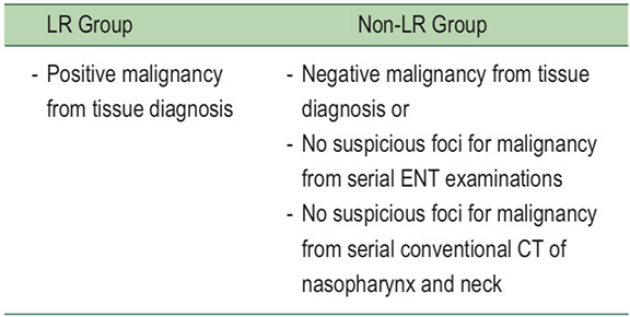
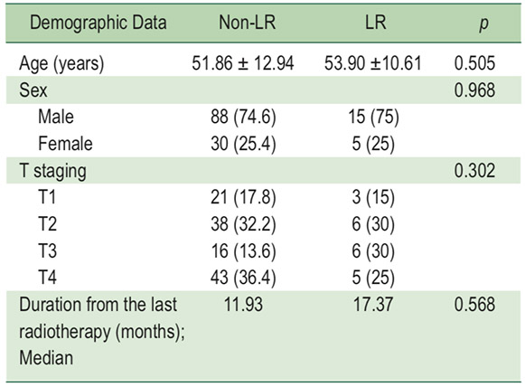
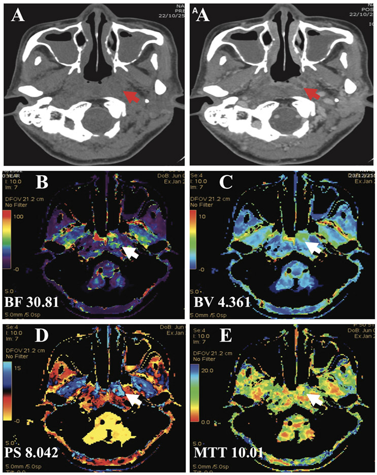
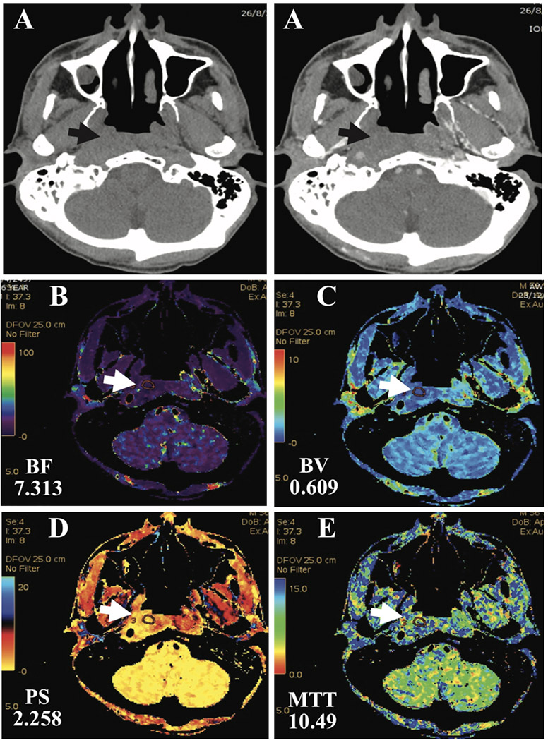
Over three years (from June 2010 to May 2013), 138 post-radiotherapy NPC patients were recruited for this prospective study. Patients were informed and consent was obtained for PCT of the nasopharynx additional to the routine contrast-enhanced CT scan of the nasopharynx. Two years follow-up after perfusion CT was performed, patients were divided into LR and non-LR groups. The perfusion CT parameters of the nasopharynx included, blood flow (BF), blood volume (BV), mean transit time (MTT) and permeability surface area product (PS). These parameters were analyzed and compared between the two groups.
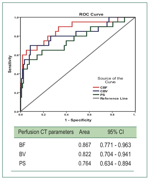
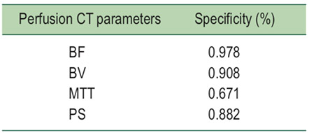
There are significantly higher PCT parameters including: BF, BV, PS and significantly lower MTT (p < 0.05) in the LR group when compared to the non-LR groups and BF and BV also demonstrated high inter-observer agreement (ICC > 0.75) between two reviewers.
PCT is a useful method to identify local recurrent/residual tumor after radiotherapy in patients with NPC.
perfNusion CT, nasopharyngeal carcinoma, local recurrence, prospective study
Received: October 30, 2018
Revision received: November 06, 2018
Accepted after revision: January 24, 2019
BKK Med J 2019;15(1): 19-27.
DOI: 10.31524/bkkmedj.2019.02.004