Electronic ISSN 2287-0237

VOLUME

ADVANCED CORONARY ATHEROSCLEROSIS AND FATAL MYOCARDIAL INFARCTION IN MILD DYSLIPIDEMIC, LOW RISK YOUNG MAN: A CASE REPORT WITH LITERATURE REVIEWED

FEBRUARY 2019 - VOL.15 | CASE REPORT

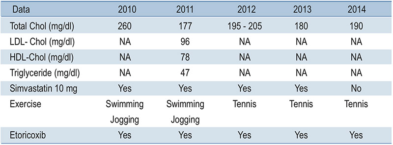
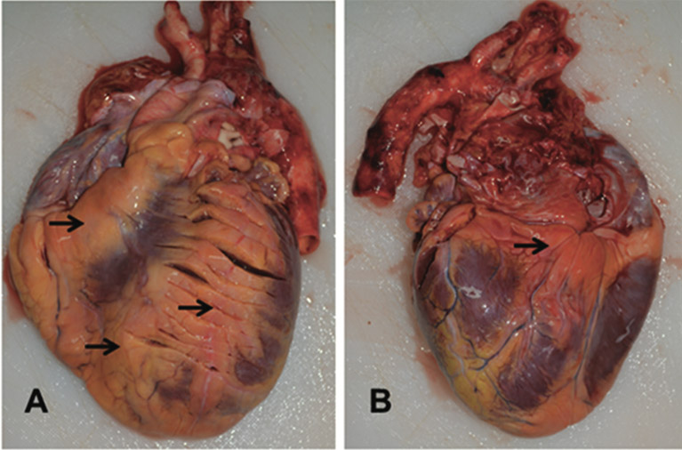
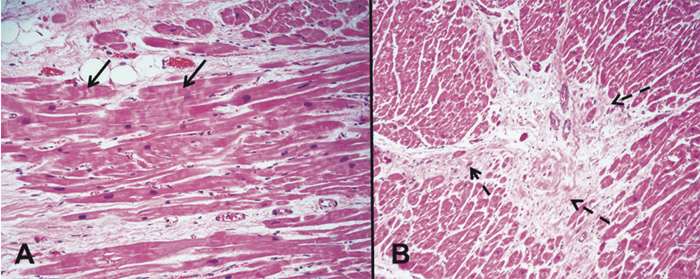
Acute Myocardial infarction (AMI) at a young age (below 45 years) is rare anddifficult to predict. We reported a fatal myocardial infarction from advanced atherosclerosisin a healthy young man who had no other major coronary risk factors except mildhypercholesterolemia. Thus, all available systemic risk scores identified him as a low riskcandidate for developing a cardiovascular event. Autopsy revealed advanced atherosclerosisin all three major coronary arteries causing acute and old myocardial infarction. Thickepicardial adipose tissue and myocardial bridging of the mid left anterior descendingartery were also noted. He frequently used etoricoxib to treat knee and back pain forconsecutive five years. Potential mechanisms of sudden death from atherosclerosis, myocardialbridging, epicardial adipose tissue and selective COXIB are discussed in moredetail below.

Keywords:

sudden death, young myocardial infarction, premature atherosclerosis, mild hypercholesterolemia, myocardial bridging, epicardial adipose tissue, COX 2 inhibitor

Received: January 28, 2019

Revision received: January 28, 2019

Accepted after revision: January 31, 2019

BKK Med J 2019;15(1): 66-72.

DOI: 10.31524/bkkmedj.2019.02.012

MEDIA
Table 1: Summary of all available lipid profiles, exercise and medication from 2010-2014
Figure 1: Pre-operative ECG in 2011 showed sinus rhythm with significant Q wave in lead III. Relative tall T wave was noted in V2 and V3. The QTc was within normal range.
Figure 2: The heart weight was above normal range, 425 gm. It was covered by the thick yellowish epicardial adipose tissue (EAT, black arrow), mostly over anterior (A), apico-basal parts of the left ventricle (B).
Figure 3: The lumen of the proximal left anterior descending artery (LAD) was occupied by atherosclerotic plaque (white arrow, A) causing over 90% luminal stenosis, as shown in histological section B. The mid LAD embedded, 10 mm deep into myocardium (brid
Figure 4: Cross section profile of the circumflex artery showed severe eccentric atheroma causing 80% luminal stenosis (A). The right coronary artery had concentric plaque with 50-80% luminal stenosis along its course (B).
Figure 5: A dark red, cross striation (black arrow, A) of myocytes, (contraction band necrosis) was noted. Area of fibrotic scar (dashed black arrow) of previous myocardial infarction was shown in B.
SEARCH VOLUME & NUMBER
GO
RECENT VOLUME & NUMBER