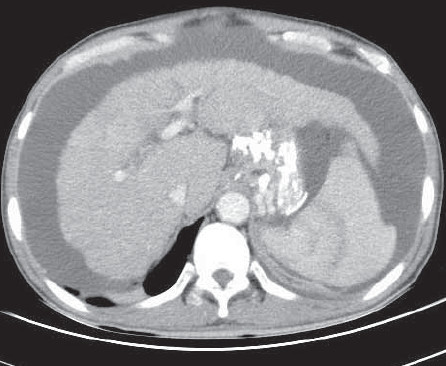
Figure 1: CT of the whole abdomen shows liver cirrhosis with multiple gastric varices post multiple glue injections.
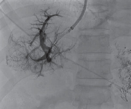
Figure 2: Transjugular to the right atrium, the catheter tip is in the right middle hepatic vein. Well demonstrated right middle hepatic vein and its distribution.
Figure 3: A. Needle punctured the hepatic vein and tipped in branch of portal vein. Venogram of intrahepatic portal veins are well visualized. B. The guide wire passed through the catheter under fluoroscopy. The guide wire passed into the portal vein.
C. The intraluminal stent is inserted then the catheter and distal end of the stent is in the portal vein. The proximal end is in the right middle hepatic vein. D. The 12 mm diameter stent is deployed in the proper position after balloon angioplasty was done.
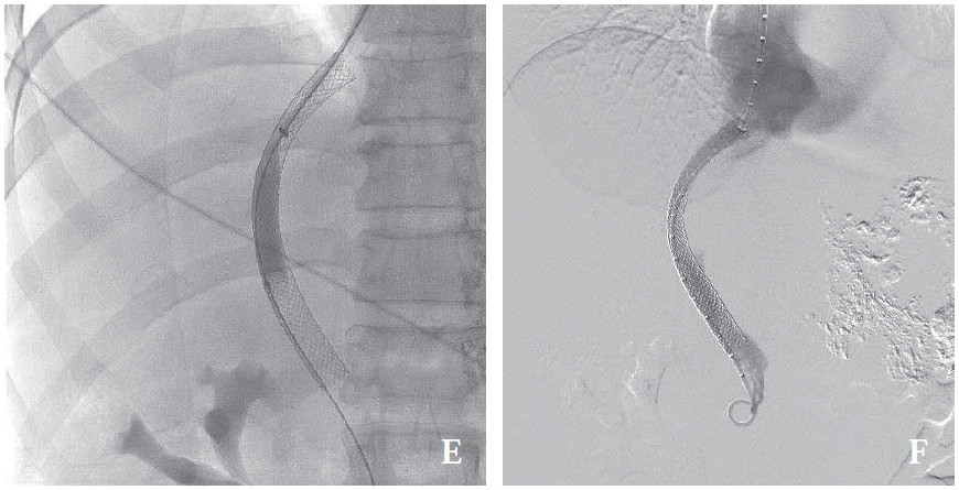
Figure 3: E. The stent is again expanded using 10 mm balloon dilatation. F. The contrast of the whole stent is well visualized. The contrast flows through the portal vein with well opacities in the right atrium.