Electronic ISSN 2287-0237

VOLUME

NEUROMYELITIS OPTICA (NMO)

SEPTEMBER 2014 - VOL.8 | CASE REPORT

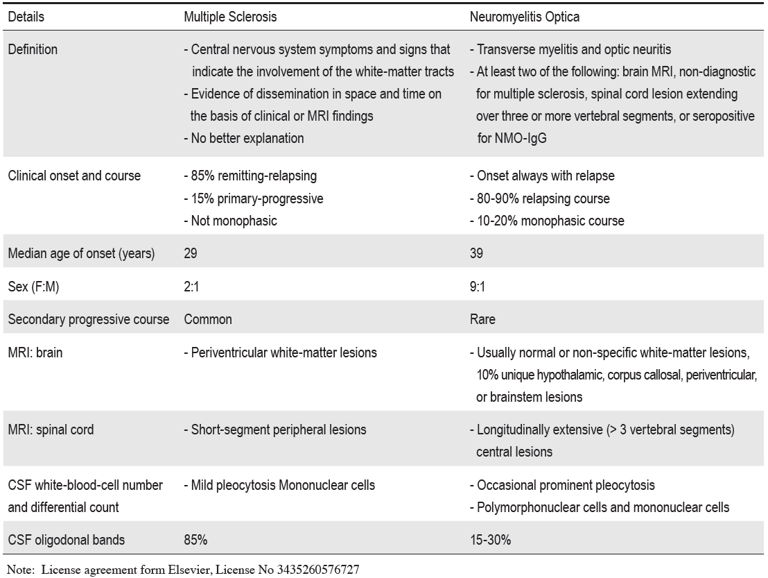
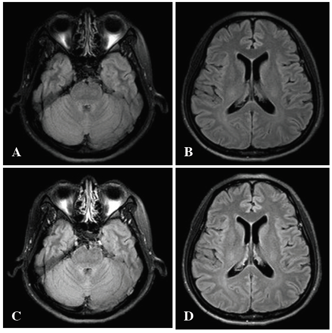
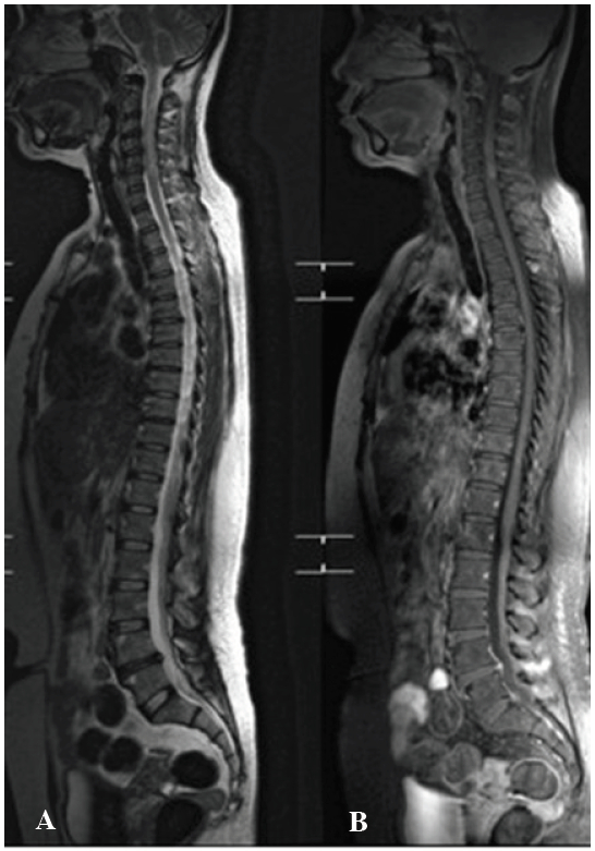
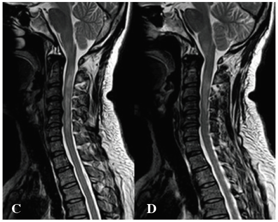
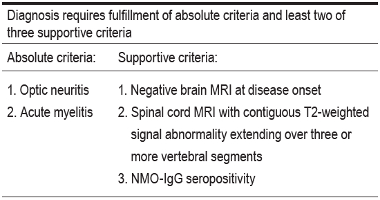
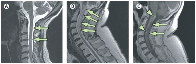
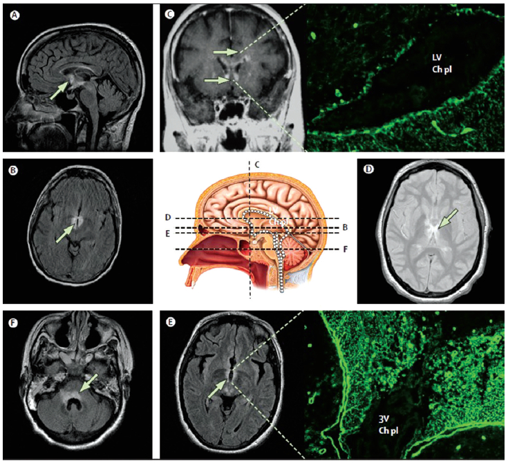
NMO is a is a rare entity which involves the central nervous system acting as an  inflammatory process by attacking the optic nerve (ON) and longitudinally  extensive transverse myelitis  (LETM). The specificity of this disease is antibody aquaporin-4 (AQP4). Repeated relapses  of the disease can lead to severe disability and blindness. MRI is none specific but at the cervical spinal cord shows the long extensive contrast enhancement. The specific diagnosis is NMO IgG.

Keywords:

neuromyelitis optica, NMO-IgG, aquaporin-4 antibody, APQ-4, multiple sclerosis, MS, oligoclonal band, azatrioprine

DOI

10.31524/bkkmedj.2014.09.007

MEDIA
Table 1:
Definition and characteristic of Multiple Sclerosis (MS) and Neuromyelitis Optica (NMO).1
Figure 1:
MRI brain T2 flair technique shows normal brain parenchyma (A,B) and post gadolinium injection reveals no enhancement (C,D).
Figure 2:
A, B: MRI whole spine neutral position: the sagittal T2- weight MR image shows a long segment of hyperintensity at cervical cord at level 2-6 with cord swelling.
Figure 2:
C, D: M R i mage s aggital T 2-weight w ith g adolinium injection: Heterogeneous patchy enhancement and swelling of the cervical spinal cord at C2-C 6 levels, suspected transverse myelitis.
Table 2:
Revised neuromyelitis optica diagnosis criteria (2006).4,5
Figure 3:
Spinal cord MRI in multiple sclerosis and neuromyelitis optica
Figure 4:
Brain lesions typical of neuromyelitis optica localised at the sites where aquaporin 4 expressions are normally highest. Representative MRI of three patients who are seropositive for NMO-IgG. The images show lesions in the periependymal regions of the brain; these sites are enriched with aquaporin 4 (white dots on centre picture of midline sagittal section). In the centre picture the dashed black lines show the anatomical level of MRI in the diagram; arrows show abnormality on fluid-attenuated inversion recovery (FLAIR), T2-weighted signal or after being given gadolinium. Patient 2 (image C; coronal, post-contrast T1-weighted image) has subependymal enhancement along the frontal horns bilaterally and in the adjacent white matter. The immunofluorescence photomicrograph linked to image C shows the binding pattern of the serum IgG from a patient with neuromyelitis optica in a mouse brain (400x). Intense immunoreactivity of basolateral ependymal cell membranes lining the lateral ventricle (LV) and extending into the subependymal astrocytic mesh coincides with aquaporin 4 immunoreactivity; the choroid plexus (Ch pl) is unstained. Patient 3 has contiguous signal abnormality throughout the periventricular tissues; diencephalon (image D; axial T2-weighted), third ventricle (image E; axial, FLAIR), and 4th ventricle (image F; axial FLAIR). Immunofluorescence photomicrograph linked to image E shows the binding pattern of the serum IgG from a patient with neuromyelitis optica in a mouse brain (400x), with intense staining of periventricular tissues (3rd ventricle, 3V); choroid plexus (Ch pl) is unstained image C is courtesy of Allen Aksamit, Mayo Clinic College of Medicine.
SEARCH VOLUME & NUMBER
GO
RECENT VOLUME & NUMBER