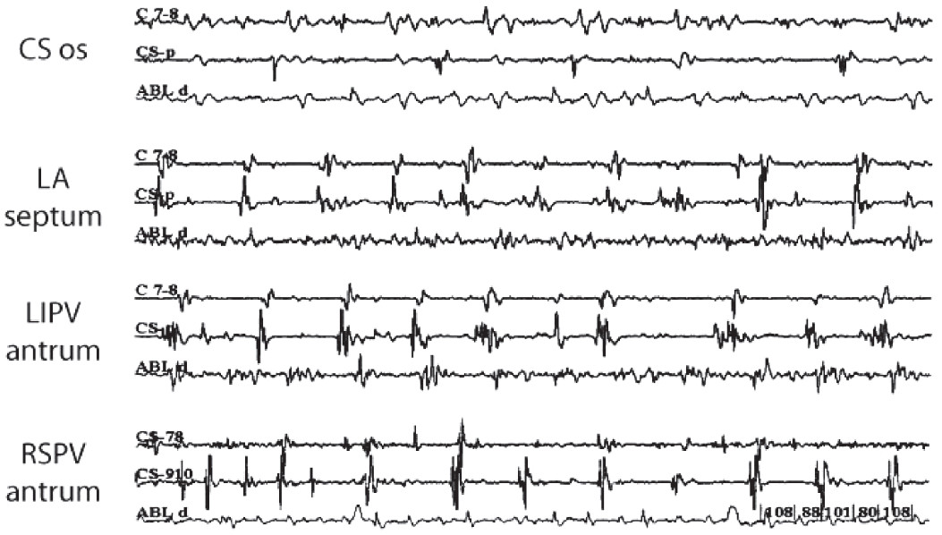
Figure 1: Various examples of CFAE that were recorded from the ablation catheter (ABL d) from different sites. Four trace panels show CFAE recorded from CS ostium (CS os), LA septum, LIPV antrum and RIPV antrum,
Each panel also shows recordings from reference site in the proximal CS (CS-7, 8 and CS-9, 10 [CSp]). The most highly fractionated electrograms can be seen in this example to exist on the LA septal wall, LIPV antrum and at the RSPV antrum.
CS = coronary sinus,
LA = left atrium,
LIPV = left anterior pulmonary vein,
RSPV = right superior pulmonary vein
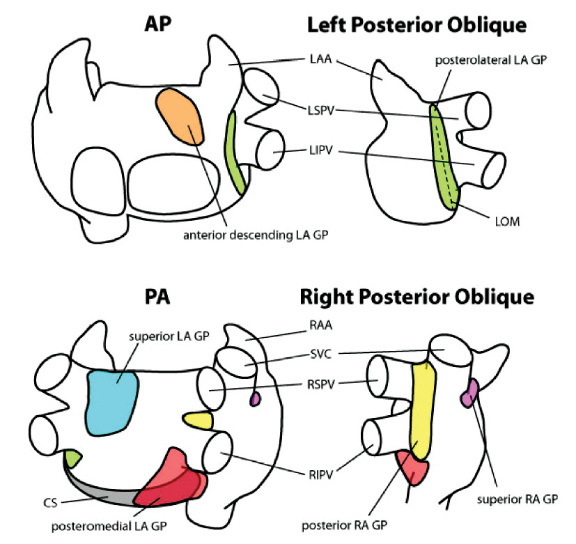
Figure 2: Six cardiac ganglionic plexi (GP) are located on or near the left and right atria and have been shown to exhibit influence on the initiation and perpetuation of AF:
superior left atrial GP, posterolateral left atrial GP, posteromedial left atrial GP, left anterior descending GP, posterior right atrial GP, and superior right atrial GP.
AP = anteroposterior, PA = posterior-anterior, LAA = left atrial appendage, RAA = right atrial appendage, CS = coronary sinus, LOM = ligament of Marshall, SVC = superior vena cava, LSPV = left superior pulmonary vein, LIPV = left inferior pulmonary vein, RSPV = right superior pulmonary vein, RIPV = right inferior pulmonary vein
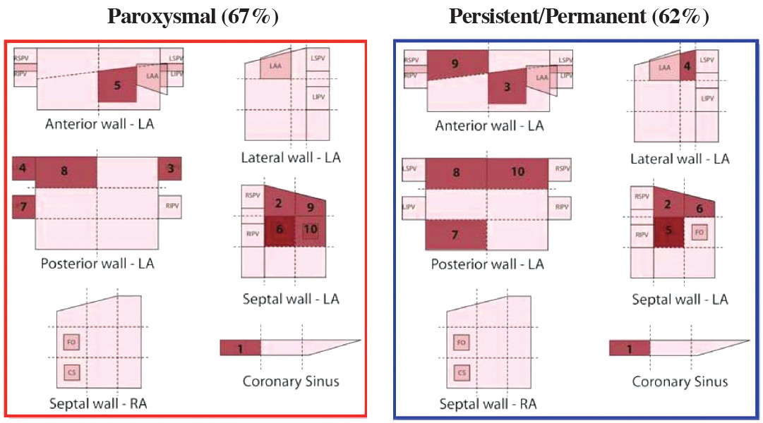
Figure 3: The most common locations of CFAE were identified (darkest shading) on a grid representing the regions of the right and left atria.
LA = left atrium, LAA = left atrial appendage, RA = right atrium, CS = coronary sinus, FO = fossa ovalis, LSPV = left superior pulmonary vein, LIPV = left inferior pulmonary vein, RSPV = right superior pulmonary vein, RIPV = right inferior pulmonary vein