Electronic ISSN 2287-0237

VOLUME

GIANT COMPLEX ODONTOMA: A CASE REPORT

FEBRUARY 2015 - VOL.9 | CASE REPORT

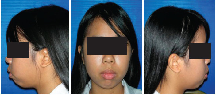
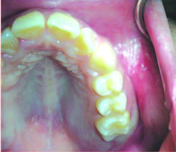
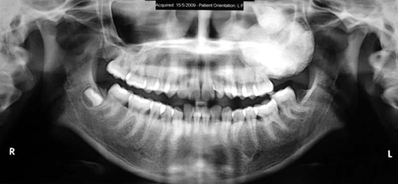
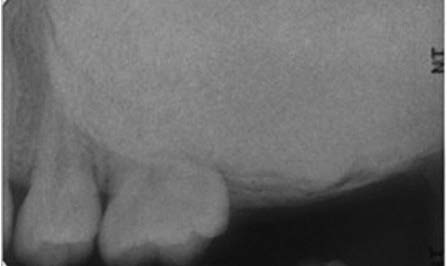
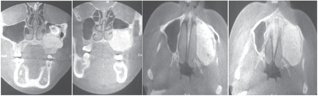
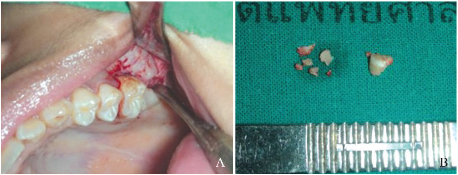
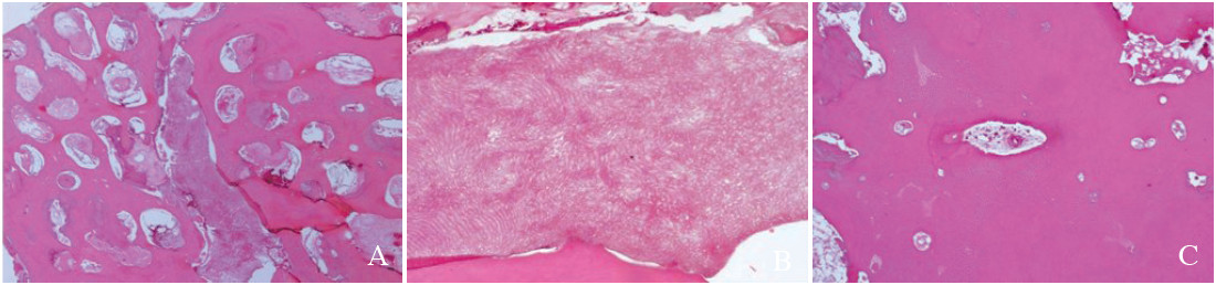
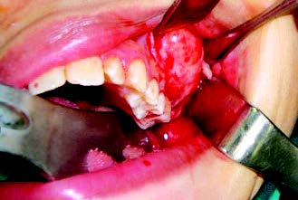
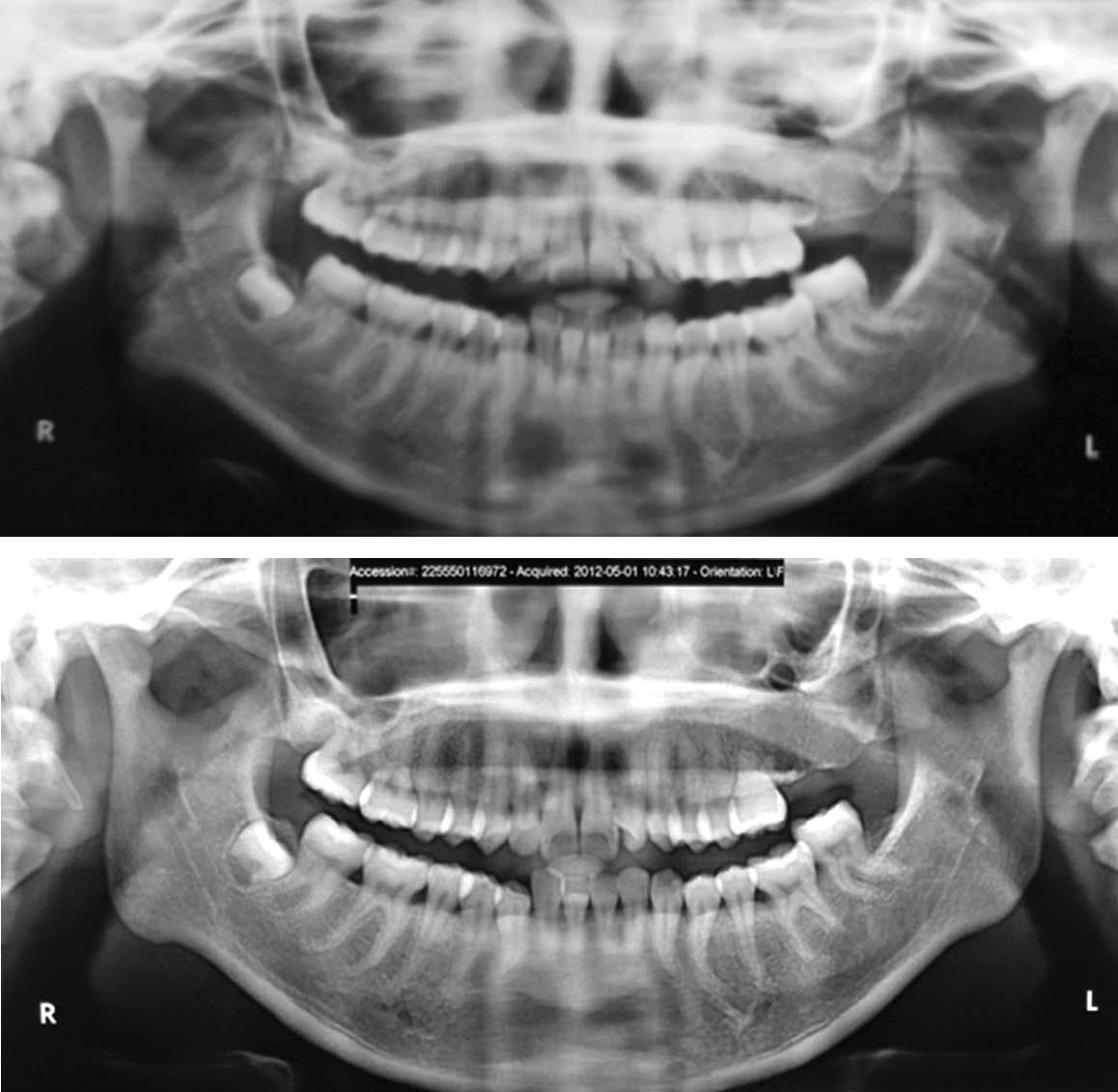
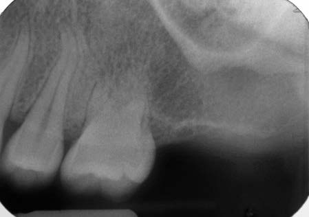
An unusual case of a large complex odontoma associated with unerupted maxillary molar of a 16-year-old Thai female patient is reported. This patient had no symptoms apart from a non-remarkable swelling at the left upper jaw bone. The lesion was accidentally detected during a radiographic investigation prior to orthodontic treatment. Upon clinical examination, the left upper second and third molars were absent. She had a bony hard swelling on the buccal and palatal sides of the left maxillary region without tenderness. The overlying mucosa appeared normal. The radiographic study revealed  a homogeneous radiopaque mass at the left maxilla involving the maxillary sinus with a displaced molar tooth. This lesion was surgically removed after the histopathologic report confirmed it to be an odontoma. The result of treatment was satisfactory at the following check-up. The details of the treatment of this odontogenic tumor, and the literature reviewed, are discussed.

Keywords:

complex odontoma, maxillary sinus, unerupted tooth

10.31524/bkkmedj.2015.02.011

MEDIA
Figure 1:
16-year-old female showed no extraoral swelling. The overlying skin appeared normal.
Figure 2:
Bone expansion of the left posterior maxilla from the second premolar to tuberosity was detected, at both the buccal and palatal side. The upper left second and third molars were absent. The overlying gingiva and other teeth in the affected region appeared normal.
Figure 3:
Panoramic radiograph showed a well-defined, unilocular homogenous radiopaque lesion, surrounded by thin radiolucent rim. The lesion extended from the left maxillary second premolar to the tuberosity and from the alveolar crest to the left maxillary sinus The root of maxillary first molar seemed to be involved. The maxillary second molar was displaced nearly to floor of orbit. The left maxil- lary third molar could not be detected.
Figure 4:
The periapical radiograph showed a radi- opaque lesion with a well-defined margin and a thin radiolucent rim. The root of the left maxillary second premolar had no resorption with a normal lamina dura and PDL space. However, the root of the left maxillary first molar could not be detected.
Figure 5:
Coronal and axial dental computed tomography showed a well-defined, homogenous radiopaque mass, measuring 3.0x3.0x3.5 cm3 inside the left maxillary alveolar process. The floor of the left maxillary sinus was elevated and the maxillary second molar was displaced superiorly. The mass expanded antero-posteriorly and bucco-palatally, causing a slight deviation of the left lateral nasal wall and perforation of the buccal alveolar bone plate.
Figure 6:
(A) An incisional biopsy was performed in the buccal area, starting from the distal of the left maxillary first molar. (B) The surgical specimen, consisting of several pieces of whitish-yellow hard mass resembling tooth structure, was sent for histological study.
Figure 7:
Histological section
Figure 8:
A full-thickness trapezoid mucoperiosteal flap was reflected to expose the mass.
Figure 9:
A thin layer of overlying bone was removed, revealing the odontoma.
Figure 10:
The mass was completely removed in multiple pieces, together with the left maxillary second molar.
Figure 11:
Radiographic examination at the 1st year (A) and 3rd year (B) after odontoma removal did not reveal any signs of recurrence.
Figure 12:
Periapical radiograph at the 3rd year after odontoma removal showed normal root of the left second premolar. However, the first molar seemed to have dental pulp obliteration, short roots and lost its lamina dura.
SEARCH VOLUME & NUMBER
GO
RECENT VOLUME & NUMBER