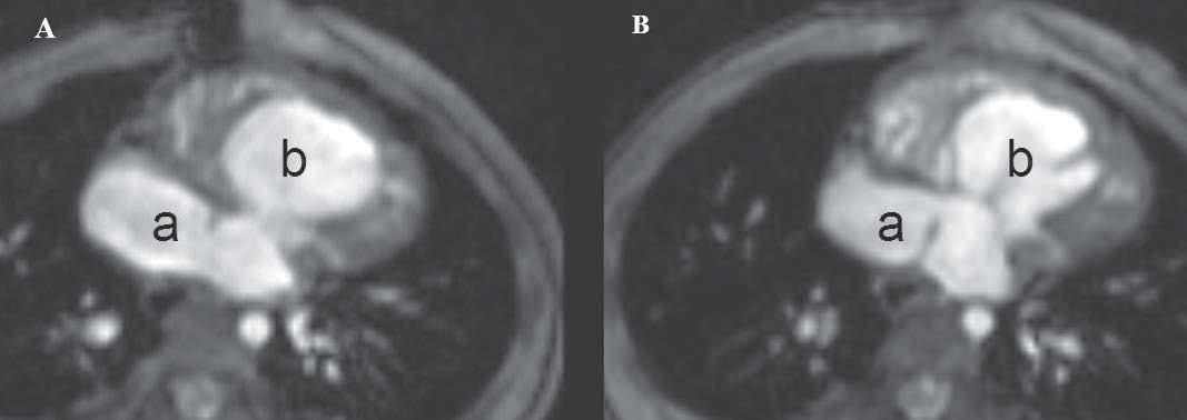
Type of total anomalous pulmonary venous return (TAPVR).
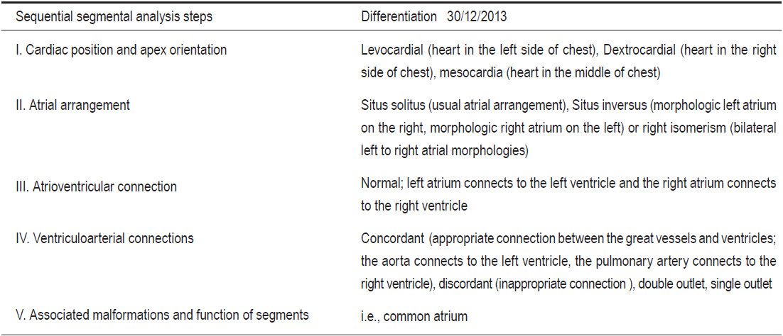
Figure 1:
Chest x-ray image on PA-supine view and magnetic resonance imaging (MRI) using gradient echo CINE pulse sequence on coronal view show the Snowman sign of TAPVR supra-cardiac type.
1A: Snowman sign on chest x-ray image. 1B: Demonstration of the components of the snowman sign on a gradient echo CINE MRI image on the coronal view, 1 = Dilated right SVC, 2 = Dilated vertical vein, 3 = Dilated left SVC, 4 = Dilated right atrium. 1C: H = Head of snowman, B = Body of snowman.
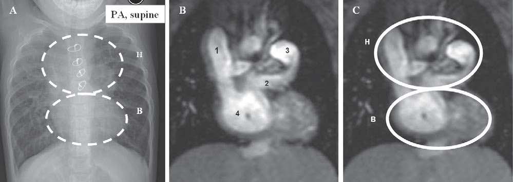
Figure 2:
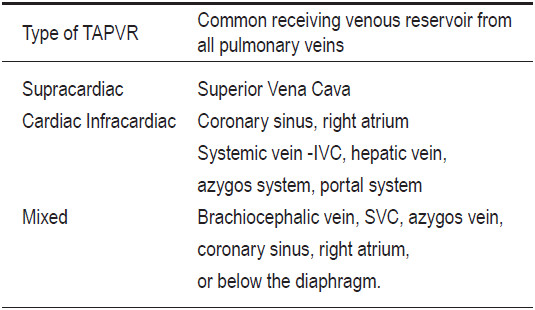
Sequential segmental demonstration approach steps I, II, III (see Table 1) are applied to MRI with a gradient echo CINE pulse sequence in an axial view. A and B show: mesocardia, common right atrium morphology (see atrial appendage character) (a) and common right morphology right ventricle (b), concordance of atrio- ventricular connection, right isomerism (bilateral atrial morphology).
Table 2:
Type of TAPVR
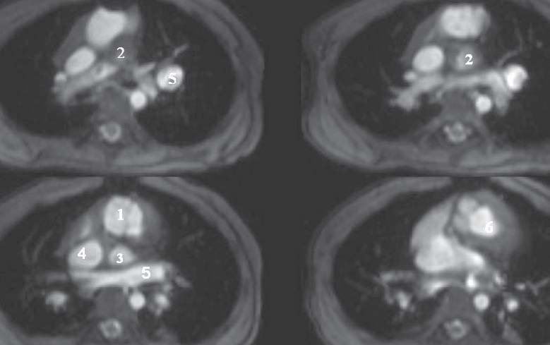
Figure 3:
Sequential segmental demonstration approach step IV is applied to an MRI with a gradient echo pulse sequence in an axial view. A-D show concordant ventriculoarterial connection, with double outlet morphology. 1 = Aortic valve, 2 = Pulmonic valve, 3 = Pulmonary viens, 4 = Superior vena cava, 5 = Vertical vein (confluence of four pulmonary arteries).
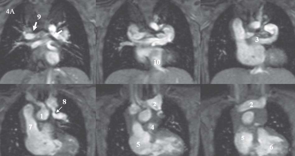
Figure 4A:
Sequential segmental demonstration approach step IV is applied on a gradient echo MRI on the coronal view: 1 = Ascending aorta, 2 = Innominate vein, 3 = Vertical vein, 4 = MPA, 5 = Right atrium, 6 = Right ventricle, 7 = Right superior vena cava, 8 = Stenosis of the connection between the vertical vein and innominate vein, 9 = Bilateral right sided tracheal line (see arrow), 10 = Inferior vena cava.
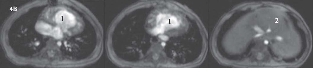
Figure 4B:
Situs ambiguous with asplenia demonstrated on gradient CINE MRI images.
1= Single ventricle, 2= Liver at the mid abdomen.
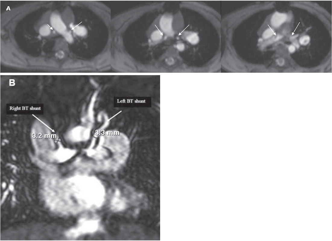
Figure 5:
A = Gradient echo CINE MRI on an axial view shows the lumen of a Blalock Taussig (BT) shunt in a short axis view.
B = MR angiography with a gadolinium contrast injection image on a coronal view which shows the left and right BT shunt.